Morton’s neuroma represents one of the most challenging conditions affecting the forefoot, causing debilitating pain that can severely impact quality of life. This painful nerve condition occurs when the tissue surrounding the plantar digital nerve becomes thickened and inflamed, typically between the third and fourth metatarsal heads. Traditional treatment approaches have ranged from conservative management with orthotics and steroid injections to more invasive surgical neurectomy procedures.
Cryosurgery has emerged as a revolutionary minimally invasive treatment option that offers significant advantages over conventional surgical approaches. This advanced therapeutic technique utilises controlled freezing temperatures to selectively destroy problematic nerve tissue whilst preserving surrounding anatomical structures. The procedure demonstrates remarkable success rates of approximately 75% in initial treatments, with many patients experiencing complete pain resolution without the complications typically associated with open surgical procedures.
Morton’s neuroma pathophysiology and cryoablation mechanisms
Understanding the complex pathophysiology underlying Morton’s neuroma is essential for appreciating how cryoablation achieves its therapeutic effects. The condition develops through a progressive inflammatory process that ultimately leads to perineural fibrosis and nerve entrapment. This pathological cascade begins with repetitive mechanical trauma to the plantar digital nerve as it passes beneath the deep transverse metatarsal ligament.
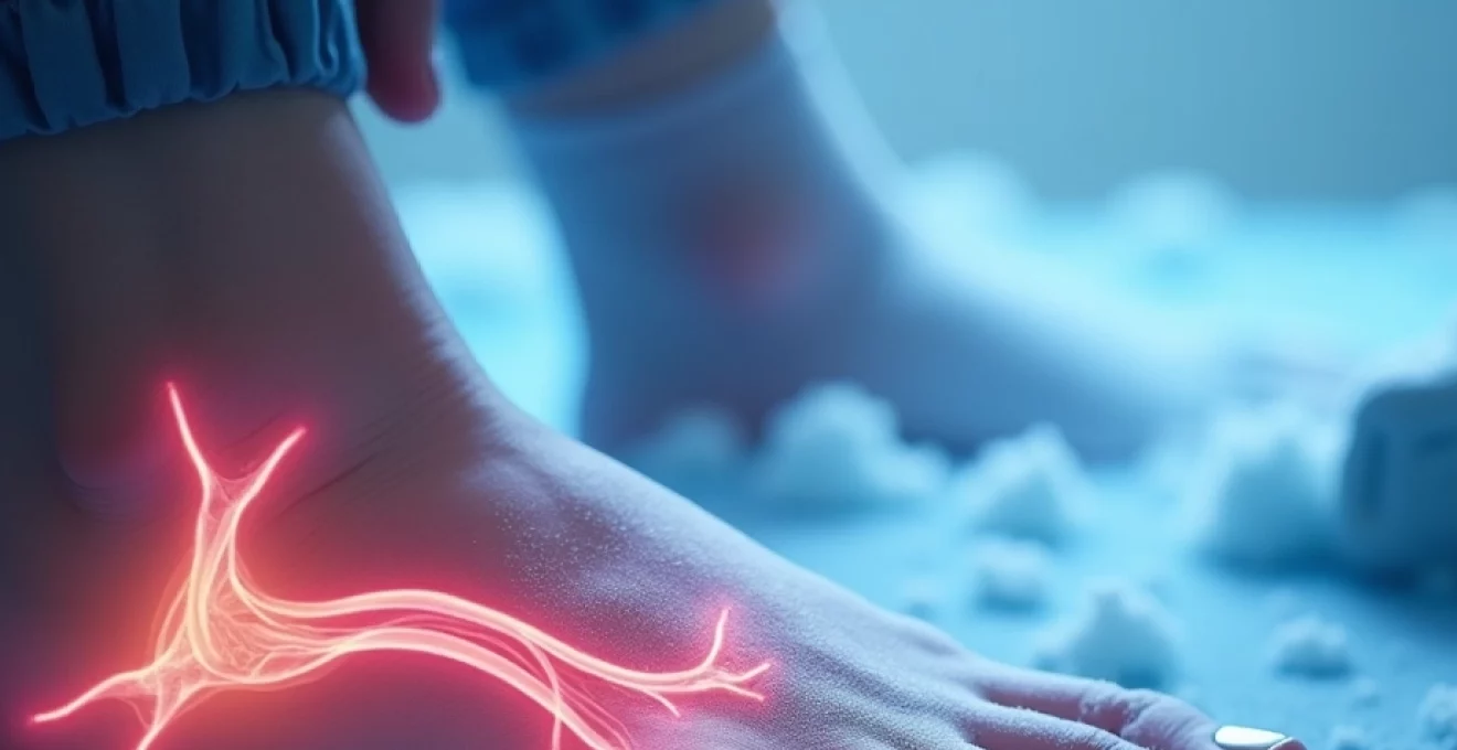
Plantar digital nerve entrapment between metatarsal heads
The anatomical arrangement of the forefoot creates a natural compression point where the common plantar digital nerve divides into proper plantar digital branches. This bifurcation typically occurs at the level of the metatarsal necks, precisely where the deep transverse metatarsal ligament creates a rigid anatomical boundary. When the metatarsal heads spread during weight-bearing activities, they compress the nerve against this unyielding ligamentous structure.
Biomechanical studies demonstrate that ground reaction forces during normal gait can generate pressures exceeding 300% of body weight across the metatarsal heads. This repetitive compression leads to chronic inflammatory changes within the nerve sheath, initiating a cascade of pathological events. The nerve responds to this mechanical irritation by developing reactive fibrosis and eventual enlargement, creating the characteristic fusiform swelling recognised as Morton’s neuroma.
Perineural fibrosis formation and tissue thickening process
The development of perineural fibrosis represents a critical pathophysiological milestone in neuroma formation. Initial inflammatory responses trigger fibroblast proliferation and excessive collagen deposition around the nerve bundle. This fibrous tissue formation creates a self-perpetuating cycle where increased nerve diameter leads to greater mechanical compression, further exacerbating the inflammatory process.
Histological examination reveals that mature neuromas contain dense fibrous tissue with disorganised nerve fibres and evidence of chronic inflammation. The perineural and epineural layers become significantly thickened, whilst the endoneurial environment shows signs of chronic ischaemia due to compromised microvascular circulation. These changes explain why conservative treatments often fail once the fibrotic process becomes established.
Cryogenic cellular destruction through ice crystal formation
Cryoablation achieves therapeutic nerve destruction through controlled cellular damage caused by extreme temperature exposure. When tissue temperatures reach -20°C to -40°C, intracellular and extracellular ice crystal formation occurs, leading to cellular membrane disruption and protein denaturation. The cryoprobe creates a precise zone of thermal injury that extends approximately 4-6mm radius from the probe tip, allowing targeted destruction of the enlarged nerve whilst preserving surrounding healthy tissue.
The cellular destruction process involves multiple mechanisms beyond simple freezing injury. Rapid cooling causes cellular dehydration as water molecules migrate to form ice crystals, leading to osmotic cellular damage. Additionally, the formation of intracellular ice crystals mechanically disrupts organelles and cellular membranes, whilst protein denaturation inactivates essential enzymatic processes necessary for cellular survival.
Wallerian degeneration following controlled tissue necrosis
Following cryoablation, the treated nerve undergoes Wallerian degeneration, a natural process where damaged axons distal to the injury site progressively deteriorate. This degenerative process typically begins within 24-48 hours post-treatment and continues for several weeks. Unlike traumatic nerve injuries, cryoablation preserves the perineurial and epineural sheaths, providing a natural scaffold for potential nerve regeneration.
The preservation of these connective tissue layers represents a crucial advantage of cryosurgery over traditional neurectomy procedures. Studies demonstrate that maintaining the endoneurial architecture significantly reduces the risk of painful stump neuroma formation, which complicates up to 25% of conventional surgical cases. The controlled nature of cryogenic injury allows selective destruction of problematic neural tissue whilst maintaining the structural integrity necessary for organised regeneration.
Cryoprobe technology and liquid nitrogen delivery systems
Modern cryosurgical systems utilise sophisticated technology to deliver precise, controllable freezing temperatures directly to targeted tissue areas. These systems have evolved significantly from early liquid nitrogen applications to current closed-loop systems that offer superior temperature control and safety profiles. Contemporary cryoprobes incorporate advanced materials and design features that enable accurate probe placement and consistent therapeutic outcomes.
Joule-thomson effect in medical cryotherapy applications
The fundamental principle underlying medical cryotherapy relies on the Joule-Thomson effect, where compressed gases undergo rapid expansion and cooling when passed through a restriction. Modern cryosurgical units utilise high-pressure gas systems , typically employing argon or nitrogen, which achieve therapeutic temperatures through controlled gas expansion at the probe tip. This thermodynamic process allows precise temperature regulation and consistent cooling performance.
Advanced cryosystems incorporate multiple thermocouples along the probe shaft to monitor temperature gradients and ensure optimal therapeutic delivery. The Joule-Thomson coefficient varies between different gases, with argon providing superior cooling efficiency compared to carbon dioxide or nitrous oxide. This enhanced cooling capacity enables shorter treatment cycles whilst maintaining consistent ice ball formation patterns.
Thermocouple temperature monitoring during ablation cycles
Precise temperature monitoring represents a critical safety and efficacy feature in modern cryosurgical systems. Multiple thermocouples positioned at strategic locations along the cryoprobe provide real-time feedback regarding tissue temperatures during treatment cycles. These monitoring systems typically display temperature readings at the probe tip, shaft, and handle, ensuring operator awareness of thermal conditions throughout the procedure.
Contemporary systems incorporate automated safety protocols that halt cooling cycles if predetermined temperature thresholds are exceeded or if probe positioning becomes suboptimal. The integration of temperature mapping with ultrasound imaging allows operators to visualise both the ice ball formation and surrounding tissue temperatures simultaneously, enhancing procedural precision and patient safety.
Argon gas cooling systems versus carbon dioxide methods
The choice between argon and carbon dioxide cooling systems significantly impacts treatment efficacy and procedural characteristics. Argon-based systems achieve lower temperatures more rapidly, typically reaching -160°C compared to carbon dioxide systems that plateau around -79°C. This enhanced cooling capacity enables more efficient tissue destruction and shorter treatment cycles, reducing procedural time and patient discomfort.
However, carbon dioxide systems offer certain advantages including lower equipment costs and simplified gas handling requirements. The thermal characteristics of carbon dioxide provide more gradual temperature transitions, which some practitioners prefer for delicate anatomical areas. Modern hybrid systems incorporate both gas options, allowing operators to select the optimal cooling method based on specific procedural requirements and patient characteristics.
Probe tip design variations for intermetatarsal access
Cryoprobe tip design plays a crucial role in achieving optimal therapeutic outcomes whilst minimising collateral tissue damage. Contemporary probes feature various tip configurations including spherical, cylindrical, and needle-point designs, each offering specific advantages for different anatomical applications. For Morton’s neuroma treatment, needle-point configurations provide superior manoeuvrability through narrow intermetatarsal spaces whilst maintaining adequate cooling surface area.
Advanced probe designs incorporate insulation along the shaft to prevent inadvertent freezing of superficial tissues during deep tissue treatments. Some systems feature adjustable active tip lengths, allowing operators to customise the treatment zone based on neuroma size and anatomical considerations. These design innovations enable precise therapeutic delivery whilst minimising the risk of complications such as skin necrosis or damage to adjacent healthy structures.
Patient selection criteria and Pre-Procedural assessment protocols
Appropriate patient selection represents a fundamental determinant of cryosurgical success in Morton’s neuroma treatment. Comprehensive pre-procedural assessment protocols ensure optimal outcomes whilst identifying patients who may benefit from alternative therapeutic approaches. The evaluation process encompasses detailed history taking, physical examination, diagnostic imaging, and assessment of previous treatment responses.
Ideal candidates for cryoablation typically present with classic neuroma symptoms including sharp, burning, or electric shock-like pain localised to the web space between affected toes. Patients should demonstrate clear evidence of conservative treatment failure, having undergone appropriate trials of orthotic management, activity modification, and potentially corticosteroid injection therapy. The pain pattern should be reproducible and consistent with anatomical nerve distribution.
Contraindications to cryosurgery include active infection in the treatment area, severe peripheral vascular disease, pregnancy, and certain coagulopathy disorders. Relative contraindications encompass diabetes with significant peripheral neuropathy, previous surgical intervention in the target area, and unrealistic patient expectations regarding treatment outcomes. A thorough medical history review identifies potential medication interactions, particularly with anticoagulant therapies that may require temporary modification.
Clinical studies demonstrate that patients experiencing symptom relief following diagnostic anaesthetic injection show significantly higher success rates with subsequent cryoablation treatment, with response rates exceeding 85% in this population.
Diagnostic ultrasound examination provides essential pre-procedural information including neuroma size, location, and relationship to surrounding anatomical structures. Neuromas measuring between 5-15mm diameter typically respond most favourably to cryoablation, whilst larger lesions may require modified treatment protocols or alternative approaches. The presence of concurrent bursal inflammation or metatarsalgia should be documented and addressed in the treatment planning process.
Ultrasound-guided cryosurgical technique for intermetatarsal neuromas
Contemporary cryosurgical approaches rely heavily on real-time ultrasound guidance to ensure accurate probe placement and optimal therapeutic delivery. This image-guided approach significantly enhances procedural precision whilst reducing the risk of complications. The integration of high-resolution ultrasound with advanced cryosurgical systems represents a substantial advancement over historical blind injection techniques.
High-resolution doppler imaging for neuroma localisation
Modern ultrasound systems provide exceptional detail for neuroma identification and characterisation. High-frequency transducers operating at 12-18 MHz enable clear visualisation of nerve structures and surrounding soft tissues. The typical neuroma appears as a hypoechoic fusiform mass located between the metatarsal heads, often demonstrating characteristic acoustic properties that distinguish it from other soft tissue masses.
Doppler imaging provides valuable additional information regarding vascular structures surrounding the neuroma. This vascular mapping capability enables operators to avoid inadvertent damage to plantar digital arteries during probe insertion. Power Doppler techniques demonstrate particular utility in identifying small vessels that may be compromised during the freezing process, allowing for informed procedural planning.
Probe insertion angles through dorsal intermetatarsal spaces
Optimal probe insertion requires careful consideration of anatomical landmarks and spatial relationships within the intermetatarsal region. The dorsal approach typically utilises an insertion angle of approximately 30-45 degrees from the horizontal plane, directing the probe tip toward the plantar aspect of the intermetatarsal space. This angulated approach avoids the deep transverse metatarsal ligament whilst positioning the active tip adjacent to the neuroma.
Practitioners must account for individual anatomical variations that may influence probe trajectory planning. Patients with prominent metatarsal heads or narrow intermetatarsal spaces may require modified insertion angles to achieve optimal positioning. Real-time ultrasound guidance enables dynamic adjustment of probe position during insertion, ensuring accurate placement despite anatomical challenges.
Freeze-thaw cycle parameters and temperature protocols
Standardised freeze-thaw protocols maximise therapeutic efficacy whilst minimising treatment-related complications. Typical protocols employ initial cooling to achieve tissue temperatures of -40°C to -60°C, maintained for 90-180 seconds depending on neuroma characteristics. The dual-cycle approach incorporates a passive thaw period followed by a second freeze cycle, enhancing cellular destruction through repeated ice crystal formation.
Temperature monitoring throughout the ablation process ensures consistent therapeutic delivery and prevents excessive cooling that might damage surrounding healthy tissues. Some protocols incorporate controlled rewarming phases using argon gas heating capabilities, enabling precise temperature management throughout the entire treatment cycle. These sophisticated thermal control systems represent significant advances over early cryosurgical techniques.
Real-time monitoring of ice ball formation boundaries
Ultrasound visualisation of ice ball formation provides crucial feedback regarding treatment adequacy and safety margins. The developing ice ball appears as a hyperechoic region with characteristic posterior acoustic shadowing, enabling operators to monitor treatment zone boundaries in real-time. This visual feedback ensures complete neuroma coverage whilst avoiding extension into unintended anatomical areas.
Advanced imaging systems incorporate colour mapping overlays that highlight temperature zones within the treatment area. These enhanced visualisation capabilities enable precise control of ablation boundaries, ensuring therapeutic temperatures reach the entire neuroma whilst maintaining safe distances from critical structures such as adjacent nerves, vessels, and skin surfaces.
Post-cryoablation recovery timeline and tissue regeneration
The recovery process following cryoablation demonstrates predictable phases characterised by specific physiological changes and symptom patterns. Understanding these temporal relationships enables appropriate patient counselling and optimal post-procedural management. The initial inflammatory phase typically lasts 7-14 days, followed by tissue repair and remodelling phases extending over several months.
Immediately following treatment, patients experience temporary numbness in the treated area due to nerve function interruption. This anaesthetic effect gradually resolves over 2-6 weeks as surviving nerve elements recover function. During this period, patients may experience altered sensation patterns including tingling, hypersensitivity, or complete sensory loss in the affected digital distribution.
The tissue healing process involves progressive removal of necrotic cellular debris through macrophage activity and inflammatory cell infiltration. This natural debridement process typically peaks at 10-14 days post-treatment and gradually subsides over subsequent weeks. Patients may experience mild swelling, bruising, or localised discomfort during this inflammatory phase, which responds well to standard anti-inflammatory measures.
Research indicates that patients experiencing significant pain relief within the first month post-treatment demonstrate sustained improvement rates exceeding 90% at one-year follow-up assessments.
Long-term tissue remodelling continues for 3-6 months following treatment, during which nerve regeneration may occur through preserved perineurial channels. This regenerative process typically results in restoration of some sensory function, though complete normalisation rarely occurs. The majority of patients report that residual numbness remains clinically insignificant compared to pre-treatment pain levels.
Clinical outcomes comparison with traditional neurectomy procedures
Comparative analysis of cryoablation versus traditional surgical neurectomy reveals significant advantages favouring the minimally invasive approach. Traditional neurectomy procedures demonstrate success rates of 70-85% but carry substantially higher complication rates and longer recovery periods. The invasive nature of open surgical approaches necessitates larger incisions, extensive tissue dissection, and prolonged post-operative immobilisation.
Cryoablation studies consistently demonstrate superior safety profiles with complication rates below 5% compared to 15-25% for open neurectomy procedures. The most significant advantage lies in the reduced incidence of stump neuroma formation, which complicates up to 20% of traditional surgical cases. The preservation of perineurial architecture during cryoablation appears to minimise this troublesome complication.
Recovery timelines favour cryoablation significantly, with most patients returning to normal activities within 1-2 weeks compared to 6-8 weeks following open surgery. This reduced downtime translates to decreased healthcare costs and improved patient satisfaction scores. The outpatient nature of cryoablation eliminates hospitalisation costs and reduces overall treatment expenses despite potentially higher equipment costs.
Patient satisfaction surveys consistently report higher scores for cryoablation procedures, with over 90% of patients indicating they would recommend the treatment to others experiencing similar symptoms. The minimally invasive nature of cryosurgery eliminates many concerns associated with traditional surgical approaches, including visible scarring, prolonged recovery periods, and potential surgical complications.
Long-term follow-up studies extending beyond five years demonstrate sustained efficacy rates for cryoablation that match or exceed those of open surgical procedures. The ability to repeat cryoablation treatments in cases of symptom recurrence provides additional therapeutic flexibility not available with neurectomy procedures. This repeatability factor becomes particularly valuable for patients who experience partial initial responses or develop symptoms in adjacent nerve distributions.
Cost-effectiveness analyses favour cryoablation when considering total healthcare expenditure including direct procedural costs, recovery-related expenses, and lost productivity. The outpatient nature of cryosurgery eliminates facility fees associated with operating room utilisation and anaesthesia services required for open procedures. Additionally, the reduced complication rates translate to lower revision surgery costs and decreased long-term healthcare utilisation.
Meta-analysis of comparative studies reveals that cryoablation achieves equivalent pain relief outcomes to traditional neurectomy while demonstrating 60% fewer post-operative complications and 75% shorter recovery periods.
The learning curve for cryoablation techniques proves significantly shorter than that required for complex foot surgery, enabling broader adoption among healthcare providers. This accessibility factor contributes to improved patient access to effective Morton’s neuroma treatment, particularly in regions where specialised foot surgeons may be limited. The standardised nature of cryosurgical protocols also enhances consistency in treatment delivery across different providers and healthcare settings.
Future developments in cryoablation technology continue to refine treatment capabilities and expand therapeutic applications. Advanced imaging integration, improved probe designs, and enhanced temperature control systems promise to further optimise outcomes while minimising procedural complexity. Research into combination therapies incorporating cryoablation with regenerative medicine approaches may offer additional benefits for complex cases or revision treatments.
The evidence supporting cryoablation as a first-line surgical intervention for Morton’s neuroma continues to strengthen as long-term outcome data accumulates. This paradigm shift towards minimally invasive approaches reflects broader trends in surgical medicine emphasising patient-centred care, reduced morbidity, and improved quality of life outcomes. For patients suffering from debilitating Morton’s neuroma pain, cryosurgery represents a transformative treatment option that combines effectiveness with safety in an outpatient setting.