Experiencing pain one week following an apicoectomy procedure raises legitimate concerns for many patients undergoing this specialised endodontic surgery. The complexity of periapical healing involves intricate biological processes that can manifest as varying degrees of discomfort during the initial recovery phase. Understanding the distinction between normal post-operative sensations and potential complications becomes crucial for patient comfort and successful treatment outcomes. Modern endodontic microsurgery techniques have significantly improved healing trajectories, yet individual responses to apical root resection can differ considerably based on numerous anatomical and physiological factors.
Understanding apicoectomy procedure and expected healing timeline
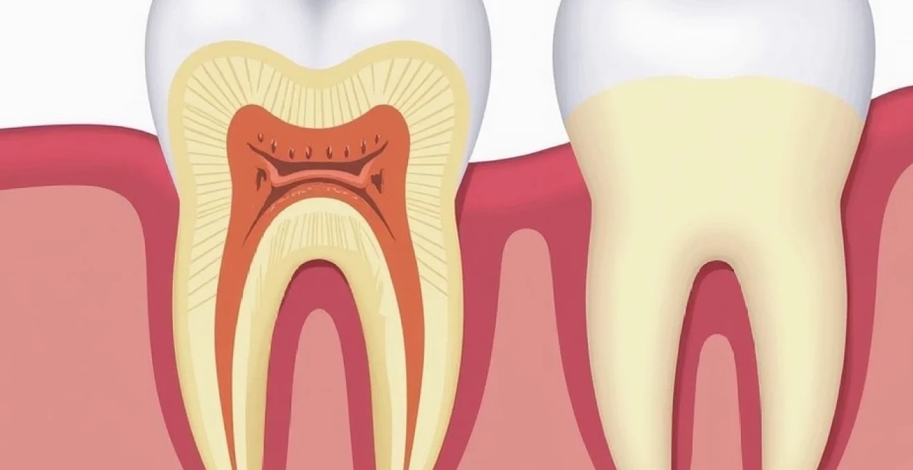
An apicoectomy represents a sophisticated retrograde endodontic procedure designed to address persistent periapical pathology when conventional root canal therapy proves insufficient. The surgical intervention involves precise removal of the infected root apex, typically 2-3 millimetres of the root tip, followed by retrograde filling with biocompatible materials. This microsurgical approach requires meticulous tissue handling and advanced visualisation techniques to ensure optimal healing outcomes.
Surgical technique: retrograde root canal therapy protocol
Contemporary apicoectomy procedures utilise operating microscopes and ultrasonic instruments to achieve unprecedented precision in root-end preparation. The surgical protocol begins with mucoperiosteal flap elevation, followed by osteotomy creation using piezoelectric instruments to minimise thermal damage. Root resection employs high-speed burs at specific angulations, typically 45 degrees to the long axis of the root, ensuring complete removal of apical ramifications and isthmuses that may harbour persistent bacteria.
Retrograde cavity preparation utilises ultrasonic tips designed specifically for root-end modification, creating a 3-millimetre deep preparation that receives mineral trioxide aggregate (MTA) or similar bioceramics. These materials provide excellent sealing properties and promote periapical healing through their biocompatibility and antimicrobial characteristics. The precision required in these procedures demands extensive specialist training and significantly influences post-operative recovery patterns.
Post-operative tissue response during first seven days
The immediate post-surgical period involves complex inflammatory cascades that generate both protective and potentially uncomfortable sensations. Vascular permeability increases substantially within the first 24-48 hours, leading to localised oedema and tissue tension that manifests as throbbing or persistent aching. This inflammatory response represents normal physiological healing rather than pathological complications, though the intensity can vary significantly between individuals.
Neutrophil migration into the surgical site peaks around day two to three post-operatively, contributing to localised discomfort through the release of inflammatory mediators. Subsequently, macrophage infiltration begins the tissue remodelling process, typically resulting in gradual pain reduction from day four onwards. Understanding this timeline helps distinguish normal healing from potential complications such as infection or delayed healing.
Periapical bone regeneration phases following apical surgery
Bone healing following apicoectomy occurs through carefully orchestrated phases that can influence pain perception throughout the first week. The initial haemostasis phase involves blood clot formation and platelet aggregation, creating a scaffold for subsequent cellular activity. This process can generate pressure-related discomfort as the clot organises within the bony cavity created during surgery.
The inflammatory phase, typically lasting 3-5 days, involves osteoclastic activity to remove damaged bone matrix alongside osteoblastic proliferation for new bone formation. This cellular activity can create sensations ranging from mild tenderness to more pronounced aching, particularly when chewing or applying pressure to the affected area. The dynamic nature of bone remodelling means that pain patterns may fluctuate rather than following a linear reduction .
Endodontic microsurgery recovery milestones
Recovery milestones following apicoectomy provide important benchmarks for assessing healing progress and identifying potential complications. Day one typically involves peak swelling and moderate discomfort, manageable with prescribed analgesics. Days two to three often represent the most challenging period, with maximum inflammatory response and potential sleep disruption due to throbbing sensations.
By day four to five, most patients experience noticeable improvement in comfort levels, though some tenderness may persist when chewing on the affected side. Day seven represents a crucial assessment point where continued moderate to severe pain may indicate complications requiring professional evaluation. The suture removal appointment, typically scheduled for this timeframe, provides an opportunity for clinical assessment of healing progress.
Normal pain parameters: week one Post-Apicoectomy assessment
Establishing normal pain parameters during the first week following apicoectomy requires understanding both subjective patient experiences and objective clinical indicators. Research demonstrates that 85-90% of patients experience their peak discomfort within the first 48 hours, with gradual improvement thereafter. However, individual variations in pain perception, healing capacity, and tissue response can create significant differences in recovery experiences.
Visual analogue scale (VAS) pain measurements day 1-7
Clinical studies utilising Visual Analogue Scale measurements provide valuable insights into expected pain trajectories following apicoectomy procedures. Day one typically registers VAS scores of 4-6 out of 10, representing moderate discomfort that may interfere with normal activities but remains manageable with appropriate analgesics. This level reflects the acute inflammatory response without necessarily indicating complications.
Days two and three often show the highest VAS scores, potentially reaching 6-8 in some patients, particularly those with complex surgical sites or multiple roots treated simultaneously. This peak represents the culmination of inflammatory mediator release and tissue oedema , creating maximum pressure and discomfort. Patients should anticipate this temporary intensification rather than interpreting it as treatment failure.
By day seven, VAS scores should demonstrate significant reduction to 2-4 range, representing mild to moderate discomfort that primarily occurs with direct pressure or chewing. Scores remaining above 6 at the one-week mark may indicate complications requiring professional assessment, though individual variations must be considered alongside other clinical factors.
Inflammatory response patterns in periradicular tissues
The periradicular tissues surrounding the surgical site undergo predictable inflammatory responses that directly correlate with pain generation. Prostaglandin E2 release peaks within 24-48 hours post-surgery, sensitising nociceptors and lowering pain thresholds in the affected area. This biochemical cascade explains why seemingly minor stimuli can produce disproportionate discomfort during the early recovery period.
Complement system activation contributes to increased vascular permeability and neutrophil chemotaxis, creating localised swelling that mechanically compresses nerve endings. The confined anatomical space within the alveolar bone intensifies this pressure effect, potentially generating deep, aching sensations that patients often describe as “pressure building up” within the jaw.
Nociceptor activation following apical root resection
Apical root resection necessarily involves transection of periodontal ligament fibres and associated nerve endings, creating immediate nociceptor activation that contributes to post-operative discomfort. These specialised sensory neurons respond to mechanical, thermal, and chemical stimuli generated during the healing process. Understanding their activation patterns helps explain why pain may seem disproportionate to the relatively minor surgical intervention.
Sensitisation of peripheral nociceptors occurs through inflammatory mediator exposure, creating hyperalgesia that can persist for several days post-operatively. This phenomenon explains why patients may experience increased sensitivity to temperature changes or pressure application even one week following surgery. The gradual resolution of sensitisation typically correlates with overall improvement in comfort levels .
Pharmacokinetics of prescribed analgesics: ibuprofen and paracetamol
Effective pain management following apicoectomy relies heavily on understanding the pharmacokinetics of commonly prescribed analgesics. Ibuprofen, a non-steroidal anti-inflammatory drug (NSAID), provides dual benefits through both analgesic and anti-inflammatory mechanisms. Peak plasma concentrations occur 1-2 hours following oral administration, with duration of action extending 4-6 hours for standard formulations.
The anti-inflammatory properties of ibuprofen specifically target cyclooxygenase enzymes responsible for prostaglandin synthesis, directly addressing the inflammatory component of post-surgical pain. Recommended dosing typically involves 400-600mg every 6-8 hours, though individual variations in metabolism and contraindications must be considered. Paracetamol provides complementary analgesic effects through different mechanisms, allowing for combination therapy when single agents prove insufficient.
Clinical studies demonstrate that combination therapy with ibuprofen and paracetamol provides superior analgesia compared to either medication alone, with reduced risk of adverse effects when used appropriately.
Distinguishing normal discomfort from complications
The ability to distinguish between expected post-operative discomfort and potential complications represents a critical skill for both patients and healthcare providers managing apicoectomy recovery. Normal healing pain typically follows predictable patterns of intensity and duration, while complications often present with distinctive characteristics that deviate from expected trajectories. Understanding these differences enables appropriate decision-making regarding when to seek professional intervention.
Normal post-apicoectomy pain demonstrates several characteristic features that differentiate it from pathological processes. The discomfort typically peaks within 48 hours and shows gradual, consistent improvement thereafter. Patients can usually identify specific triggers such as chewing, temperature changes, or direct pressure, while the pain remains localised to the surgical site without radiating to distant areas. Most importantly, normal pain responds appropriately to prescribed analgesics and follows expected pharmacokinetic patterns .
Complications, conversely, often present with pain that either fails to improve or actually worsens after the initial 48-72 hour period. Infection-related pain may demonstrate a characteristic throbbing quality accompanied by fever, malaise, or purulent discharge. Nerve injury can produce sharp, electric-like sensations or persistent numbness extending beyond the immediate surgical area. Dry socket formation, though less common in apicoectomy than extraction procedures, can create severe, radiating pain that fails to respond to conventional analgesics.
The temporal relationship between pain and healing milestones provides additional diagnostic clues for distinguishing normal from abnormal recovery patterns. Expected improvement should become apparent by day four to five, with significant reduction in spontaneous pain and increased tolerance for normal activities. Complications often disrupt this timeline, creating persistent or escalating discomfort that interferes with sleep, eating, or routine functions beyond the expected recovery period.
Pain management protocols for apicoectomy recovery
Comprehensive pain management following apicoectomy requires a multimodal approach that addresses both inflammatory and neuropathic components of post-surgical discomfort. Evidence-based protocols emphasise prevention of pain sensitisation through pre-emptive analgesia combined with sustained anti-inflammatory therapy throughout the acute healing phase. This approach recognises that effective pain control not only improves patient comfort but also promotes optimal healing outcomes by reducing stress-related complications.
Multi-modal analgesia approach: NSAIDs and opioid alternatives
Contemporary pain management protocols prioritise non-opioid alternatives that provide effective analgesia while minimising risks associated with narcotic medications. The combination of NSAIDs with paracetamol forms the foundation of most post-apicoectomy pain management regimens, offering complementary mechanisms of action that address different aspects of surgical discomfort. This approach has demonstrated superior efficacy compared to single-agent therapy while reducing the likelihood of breakthrough pain episodes.
Ibuprofen remains the NSAID of choice for most patients, providing both analgesic and anti-inflammatory benefits crucial for managing post-surgical inflammation. Typical dosing involves 400-600mg every six hours for the first 48-72 hours, followed by as-needed administration based on symptom severity. Paracetamol at 1000mg every six hours provides additional analgesic coverage through different pathways, creating synergistic effects when combined with NSAIDs.
For patients unable to tolerate NSAIDs due to gastrointestinal concerns or cardiovascular contraindications, alternative strategies include selective COX-2 inhibitors or topical analgesics applied to the affected area. These alternatives may require dosing adjustments and careful monitoring to ensure adequate pain control without compromising healing . The goal remains achieving comfortable recovery while avoiding the sedation and dependency risks associated with opioid medications.
Corticosteroid administration: prednisolone dosing regimens
Corticosteroid therapy can provide significant benefits for managing post-apicoectomy inflammation and associated discomfort, particularly in cases involving extensive surgical manipulation or patients with heightened inflammatory responses. Prednisolone represents the most commonly prescribed corticosteroid for dental surgical applications, offering potent anti-inflammatory effects with predictable pharmacokinetics and manageable side effect profiles when used short-term.
Typical prednisolone dosing regimens for apicoectomy recovery involve 25-40mg daily for 3-5 days, initiated within 24 hours of surgery for maximum effectiveness. This approach can dramatically reduce post-operative swelling and associated pressure-related pain, though individual patient factors including medical history and concurrent medications must be carefully considered. The short duration of therapy minimises risks associated with corticosteroid administration while providing maximum anti-inflammatory benefit during the critical healing period.
Cryotherapy applications for orofacial swelling reduction
Cryotherapy represents a simple yet effective adjunctive treatment for managing post-apicoectomy swelling and associated discomfort. The application of controlled cold temperatures creates vasoconstriction that reduces inflammatory exudate formation while providing temporary analgesic effects through nerve conduction modulation. Proper cryotherapy technique involves 15-20 minute applications every 2-3 hours during the first 48 hours post-surgery.
The physiological effects of cryotherapy extend beyond simple vasoconstriction to include reduced cellular metabolism and inflammatory mediator release. This creates a cascade of beneficial effects that can significantly improve patient comfort during the critical early healing period. However, excessive cold application can potentially impair healing through compromised circulation, emphasising the importance of appropriate timing and duration protocols.
Research demonstrates that patients utilising proper cryotherapy protocols experience 30-40% reduction in peak swelling compared to those relying solely on pharmacological interventions.
When to contact your endodontist: red flag symptoms
Recognising red flag symptoms that warrant immediate professional attention represents a crucial aspect of safe apicoectomy recovery. While mild to moderate discomfort during the first week falls within normal parameters, certain warning signs indicate potential complications requiring urgent intervention. Understanding these symptoms empowers patients to seek appropriate care while avoiding unnecessary anxiety about normal healing processes.
Severe, escalating pain that fails to respond to prescribed analgesics represents the most concerning red flag symptom following apicoectomy. This type of discomfort often indicates infection, nerve injury, or other complications that require immediate professional assessment. Pain that worsens progressively after day three, particularly when accompanied by fever, swelling, or discharge, suggests bacterial infection that may require antibiotic therapy or surgical intervention.
Persistent numbness or altered sensation extending beyond the immediate surgical area indicates potential nerve involvement that requires urgent evaluation. While temporary sensory changes directly adjacent to the surgical site may occur normally, extensive or progressive numbness suggests nerve injury requiring specialised management. Early intervention in nerve-related complications can significantly improve long-term outcomes and prevent permanent sensory deficits.
- Temperature elevation above 38°C (100.4°F) persisting beyond 48 hours post-surgery
- Purulent discharge or foul taste emanating from the surgical site
- Excessive bleeding that cannot be controlled with direct pressure
- Difficulty swallowing or opening the mouth progressively worsening after day two
- Signs of allergic reaction including rash, difficulty breathing, or facial swelling beyond the surgical area
The timing of symptom development provides important diagnostic information for distinguishing normal healing from pathological processes. Complications typically manifest either immediately post-surgery or after day three, while normal healing follows predictable improvement patterns. Patients should maintain detailed symptom logs documenting pain levels, medication effectiveness, and associated symptoms to provide accurate information during professional consultations.
Long-term prognosis following periapical surgery
The long-term prognosis following apicoectomy procedures has improved dramatically with advances in surgical techniques, materials, and post-operative care protocols. Contemporary success rates exceed 85-90% at five-year follow-up periods, with many treated teeth maintaining function and comfort for decades. However, the initial week of recovery plays a crucial role in determining these long-term outcomes, as proper healing during this period establishes the foundation for sustained success.
Pain experienced during the first week
typically serves as a strong predictor of overall treatment success. Research indicates that patients experiencing appropriate pain reduction by day seven demonstrate significantly higher long-term success rates compared to those with persistent severe discomfort. The correlation between early healing patterns and ultimate treatment outcomes emphasises the importance of monitoring pain levels during this critical period.
Factors influencing long-term prognosis include the patient’s overall health status, oral hygiene maintenance, and adherence to post-operative care instructions. Smoking cessation, proper wound care, and regular dental maintenance significantly improve the likelihood of sustained treatment success. Additionally, the quality of the retrograde seal and completeness of infected tissue removal during surgery directly impact long-term outcomes, highlighting the importance of selecting experienced endodontic specialists for these procedures.
Follow-up radiographic examinations at six months, one year, and annually thereafter provide objective measures of healing success through evidence of bone regeneration around the treated root apex. Patients who experience normal pain patterns during the first week typically demonstrate complete radiographic healing within 6-12 months , establishing a strong foundation for decades of continued function. The investment in proper early recovery management thus yields substantial long-term benefits for both comfort and treatment durability.
Contemporary apicoectomy techniques have transformed the prognosis for teeth that previously faced certain extraction. The combination of microsurgical precision, biocompatible materials, and evidence-based recovery protocols has created treatment outcomes that rival or exceed those of alternative tooth replacement options. Patients can expect their treated teeth to function normally for many years, provided they maintain appropriate oral hygiene and attend regular professional maintenance appointments.
While the experience of pain during the first week following apicoectomy can cause understandable concern, understanding normal healing patterns provides reassurance and guidance for appropriate recovery management. The sophisticated biological processes involved in periapical healing necessarily involve some degree of discomfort, but this temporary inconvenience typically yields excellent long-term results when managed appropriately. Most patients find that their initial concerns about post-operative pain prove unfounded when weighed against the successful preservation of their natural tooth and restoration of comfortable function.