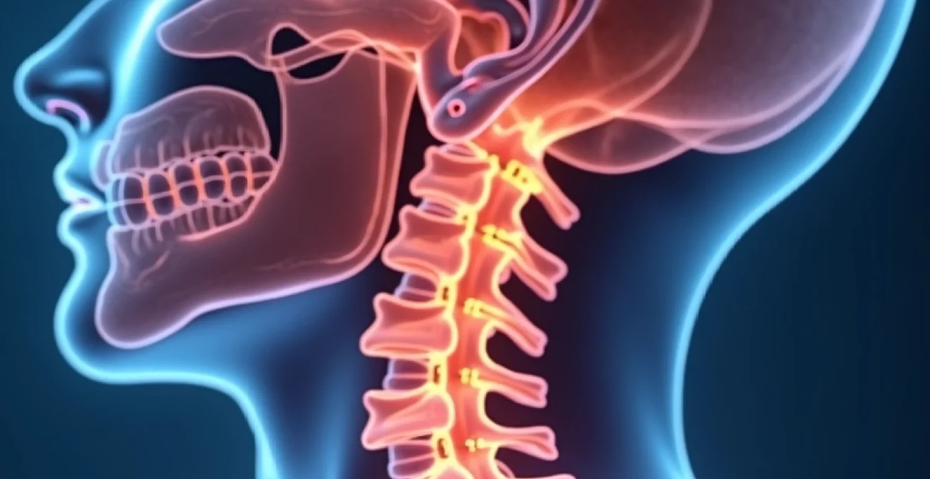
Electric shock sensations in the neck represent one of the most perplexing and distressing neurological symptoms that patients can experience. These sudden, shooting pains that radiate from the cervical spine often catch individuals off guard, creating moments of intense discomfort that can significantly impact daily activities. The phenomenon, characterised by sharp, lightning-like sensations that travel through the neck and potentially into the arms or spine, affects thousands of people worldwide and stems from various underlying conditions ranging from benign muscle tension to serious neurological disorders.
Understanding the diverse causes behind cervical electric shock sensations is crucial for both patients and healthcare providers. The complexity of the cervical region , with its intricate network of nerves, blood vessels, and musculoskeletal structures, means that pinpointing the exact source requires comprehensive evaluation and expert clinical assessment. Modern diagnostic techniques have revolutionised our ability to identify these conditions, leading to more targeted and effective treatment approaches that can restore quality of life for affected individuals.
Neurological causes of cervical electric shock sensations
Neurological disorders represent the most common category of conditions responsible for electric shock sensations in the neck region. These conditions involve direct damage or dysfunction of the nervous system, creating abnormal electrical impulses that manifest as sudden, sharp pains. The complexity of neurological causes requires careful evaluation to distinguish between central and peripheral nervous system involvement.
Cervical radiculopathy and C5-C8 nerve root compression
Cervical radiculopathy occurs when nerve roots exiting the spinal cord become compressed or irritated, typically between the C5 and C8 vertebral levels. This compression creates characteristic electric shock sensations that follow specific dermatomal patterns, allowing clinicians to identify the affected nerve root with remarkable precision. The shooting pains often extend beyond the neck , travelling down the shoulder blade, arm, and even into the fingers, depending on which nerve root is compromised.
Herniated cervical discs represent the primary cause of nerve root compression, particularly affecting individuals between ages 30 and 50. The gelatinous nucleus pulposus can protrude through the annulus fibrosus, creating direct pressure on adjacent nerve roots. This mechanical compression triggers inflammatory responses that amplify pain signals, resulting in the characteristic electric shock sensations that patients describe as unbearable.
Lhermitte’s sign in multiple sclerosis and spinal cord lesions
Lhermitte’s sign presents as a distinctive electric shock sensation that travels down the spine when the neck is flexed forward. This neurological phenomenon occurs in approximately 16-30% of individuals with multiple sclerosis, making it a significant diagnostic marker for demyelinating conditions. The sensation results from damaged myelin sheaths surrounding spinal cord nerves, which become hypersensitive to mechanical stimulation during neck movement.
Multiple sclerosis lesions in the cervical spinal cord create areas of demyelination that disrupt normal nerve conduction. When patients bend their necks forward, the mechanical stress on these damaged areas triggers abnormal electrical discharges that cascade down the spinal cord. This phenomenon can also occur in other conditions affecting spinal cord integrity, including transverse myelitis, spinal cord tumours, and radiation-induced myelopathy.
The electric shock sensation of Lhermitte’s sign serves as an important clinical indicator of spinal cord pathology, particularly when associated with other neurological symptoms such as weakness, numbness, or coordination difficulties.
Trigeminal neuralgia extension and occipital nerve involvement
Occipital neuralgia affects the greater and lesser occipital nerves that innervate the posterior scalp and upper neck region. This condition produces sharp, shooting pains that begin at the base of the skull and radiate upward over the head, often accompanied by tenderness along the nerve pathways. The electric shock quality of occipital neuralgia pain frequently extends into the cervical region, creating confusion with other neurological conditions.
Trigeminal neuralgia, while primarily affecting facial sensation, can occasionally produce referred pain patterns that extend into the upper cervical region. The anatomical connections between cranial nerves and upper cervical nerve roots create complex pain referral patterns that can manifest as electric shock sensations in the neck. Understanding these interconnections is crucial for accurate diagnosis and appropriate treatment planning.
Peripheral neuropathy affecting greater auricular and transverse cervical nerves
Peripheral neuropathy of the cervical region involves damage to superficial sensory nerves, including the greater auricular nerve and transverse cervical nerves. These conditions can result from diabetes mellitus, vitamin B12 deficiency, autoimmune disorders, or exposure to neurotoxic substances. The damaged peripheral nerves become hyperexcitable, generating spontaneous electrical discharges that patients experience as sudden shock-like sensations.
Diabetic peripheral neuropathy affects approximately 50% of individuals with long-standing diabetes and can manifest in cervical nerve distributions. The metabolic disruption of nerve function creates areas of demyelination and axonal damage that predispose to abnormal electrical activity. These neuropathic changes often progress gradually, with electric shock sensations representing an early manifestation of nerve dysfunction.
Musculoskeletal disorders triggering cervical Electroshock-Like pain
Musculoskeletal disorders of the cervical spine can create mechanical irritation of neural structures, resulting in electric shock sensations that mimic neurological conditions. These disorders involve structural abnormalities of bones, joints, ligaments, or muscles that create secondary neurological symptoms. Understanding the musculoskeletal contribution to cervical electric shock sensations is essential for comprehensive patient care.
Cervical spondylosis and foraminal stenosis at C4-C7 levels
Cervical spondylosis represents age-related degenerative changes affecting the cervical spine, with most significant changes occurring at C5-C6 and C6-C7 levels. These degenerative processes include disc height loss, osteophyte formation, and ligamentum flavum thickening, all of which contribute to neural foraminal narrowing. The progressive stenosis creates intermittent nerve compression that manifests as electric shock sensations, particularly during neck movement or specific postures.
Foraminal stenosis occurs when the bony openings through which nerve roots exit the spinal canal become narrowed. This narrowing can be static, caused by structural changes, or dynamic, occurring only during certain neck positions. The mechanical compression of nerve roots triggers inflammatory responses and abnormal electrical activity, resulting in the characteristic shooting pains that patients describe as electric shocks.
Research indicates that cervical spondylosis affects up to 85% of individuals over age 60, making it a leading cause of cervical electric shock sensations in older populations. The degenerative changes progress gradually, with symptoms often developing insidiously before reaching clinical significance. Early intervention with appropriate conservative measures can slow progression and reduce symptom severity.
Atlantoaxial instability and upper cervical dysfunction
Atlantoaxial instability involves abnormal movement between the first and second cervical vertebrae, creating potential for neural structure irritation. This condition can result from rheumatoid arthritis, congenital abnormalities, or traumatic injuries affecting the atlantoaxial joint complex. The instability creates intermittent compression of the upper cervical nerve roots and potentially the brainstem, manifesting as electric shock sensations that often accompany head and neck movement.
Upper cervical dysfunction encompasses a range of conditions affecting the occiput-atlas-axis complex, including joint restriction, muscle imbalance, and ligamentous laxity. These dysfunctions can create aberrant proprioceptive input and mechanical irritation of the upper cervical nerve roots. The resulting electric shock sensations often accompany headaches , dizziness, and neck stiffness, creating a complex symptom pattern that requires expert evaluation.
Myofascial trigger points in sternocleidomastoid and scalene muscles
Myofascial trigger points in cervical musculature can create referred pain patterns that mimic neurological electric shock sensations. The sternocleidomastoid and scalene muscles are particularly prone to trigger point development, especially in individuals with forward head posture or repetitive strain activities. These hyperirritable spots within muscle fibres can generate sudden, sharp pains that radiate in characteristic patterns.
Trigger points in the scalene muscles can create compression of the brachial plexus, resulting in neurogenic symptoms that include electric shock sensations radiating into the arm. The anatomical relationship between these muscles and neural structures creates the potential for mechanical irritation that manifests as sudden, shooting pains. Understanding these relationships is crucial for differentiating myofascial causes from primary neurological conditions.
Thoracic outlet syndrome and brachial plexus compression
Thoracic outlet syndrome involves compression of the brachial plexus nerve bundle as it passes through the thoracic outlet, creating neurological symptoms that can include electric shock sensations radiating from the neck into the arm. This condition affects approximately 8% of the population and can result from anatomical variations, trauma, or repetitive overhead activities. The compression can be dynamic , occurring only during specific arm positions or activities.
Brachial plexus compression can occur at multiple anatomical sites, including between the scalene muscles, beneath the pectoralis minor muscle, or at the costoclavicular space. Each compression site creates characteristic symptom patterns, with electric shock sensations representing a common manifestation. The diagnosis requires careful clinical evaluation and often specialised testing to identify the specific compression site.
Vascular and circulatory factors in cervical electric sensations
Vascular disorders affecting the cervical region can create electric shock sensations through several mechanisms, including nerve ischaemia, vascular compression of neural structures, and inflammatory vasculopathy. These conditions are often overlooked in the evaluation of cervical electric shock sensations but represent important diagnostic considerations that require specific treatment approaches.
Vertebrobasilar insufficiency can create transient ischaemia of neural structures in the cervical region, manifesting as sudden electric shock sensations accompanied by dizziness, visual disturbances, or balance problems. This condition typically affects older individuals with atherosclerotic disease and can be triggered by specific neck movements that compromise blood flow through the vertebral arteries. The vascular contribution to cervical symptoms requires careful evaluation with appropriate imaging studies.
Temporal arteritis and other inflammatory vasculopathies can affect blood vessels supplying cervical neural structures, creating ischaemic changes that manifest as electric shock sensations. These conditions often present with additional systemic symptoms, including headache, jaw claudication, and elevated inflammatory markers. The diagnosis requires high clinical suspicion and prompt treatment to prevent serious complications.
Cervical artery dissection represents a serious but rare cause of cervical electric shock sensations, typically occurring following trauma or sudden neck movements. The dissection can create direct compression of adjacent neural structures or compromise blood flow to nerve roots. This condition requires immediate medical attention and specialised imaging studies for accurate diagnosis and appropriate treatment.
Medication-induced and iatrogenic cervical dysesthesia
Medication-induced cervical electric shock sensations represent an important but often overlooked cause of neurological symptoms. Certain medications can affect nerve function directly or alter neurotransmitter balance, creating abnormal electrical activity that manifests as sudden shock-like sensations. Understanding these medication-related causes is crucial for comprehensive patient evaluation and management.
Selective serotonin reuptake inhibitor (SSRI) discontinuation syndrome commonly produces electric shock sensations, often described as “brain zaps” that can radiate into the cervical region. These symptoms typically occur when SSRIs are discontinued abruptly or when doses are reduced too rapidly. The mechanism involves alterations in serotonin neurotransmission that create temporary neurological instability. Gradual medication tapering can prevent or minimise these discontinuation symptoms.
Chemotherapy-induced peripheral neuropathy affects up to 68% of patients receiving neurotoxic agents such as platinum compounds, taxanes, or vinca alkaloids. These medications can damage peripheral nerves, creating areas of demyelination and axonal injury that predispose to abnormal electrical activity. The resulting electric shock sensations can persist for months or years following chemotherapy completion, requiring specialised neuropathic pain management.
Cervical spine surgical procedures can occasionally result in iatrogenic nerve injury, creating post-operative electric shock sensations. These complications can result from direct nerve trauma, inflammation, or scar tissue formation around neural structures. The incidence of significant neurological complications following cervical spine surgery ranges from 0.2% to 5%, depending on the specific procedure performed and individual patient factors.
Post-operative electric shock sensations following cervical spine surgery require careful evaluation to distinguish between expected temporary symptoms and significant complications requiring intervention.
Diagnostic imaging and electrophysiological assessment protocols
Accurate diagnosis of cervical electric shock sensations requires a systematic approach combining clinical evaluation with appropriate diagnostic testing. The complexity of potential causes necessitates a comprehensive assessment strategy that can identify both structural abnormalities and functional disturbances of the nervous system.
Cervical MRI with gadolinium enhancement for demyelinating lesions
Magnetic resonance imaging represents the gold standard for evaluating structural causes of cervical electric shock sensations. Cervical MRI can identify disc herniations, spinal stenosis, tumours, and inflammatory conditions affecting the spinal cord and nerve roots. Gadolinium enhancement improves detection of inflammatory lesions, tumours, and areas of active demyelination that may not be visible on standard sequences.
Specialised MRI sequences, including diffusion tensor imaging and magnetic resonance spectroscopy, can provide additional information about tissue integrity and metabolic activity. These advanced techniques are particularly valuable for evaluating demyelinating conditions such as multiple sclerosis, where conventional imaging may appear normal despite significant clinical symptoms. The integration of these advanced techniques into routine clinical practice continues to evolve.
Electromyography and nerve conduction studies for radiculopathy
Electromyography (EMG) and nerve conduction studies provide objective assessment of peripheral nerve function and can localise sites of nerve damage with high precision. These studies are particularly valuable for evaluating suspected cervical radiculopathy, allowing clinicians to differentiate between nerve root compression and peripheral nerve disorders. The combination of motor and sensory nerve conduction studies provides comprehensive evaluation of nerve function.
EMG testing can detect denervation changes in muscles innervated by compressed nerve roots, often before clinical weakness becomes apparent. The timing of EMG changes following nerve injury follows predictable patterns, with abnormalities typically appearing 2-3 weeks after acute compression. Serial EMG studies can monitor recovery following treatment interventions and guide decisions regarding surgical intervention.
Somatosensory evoked potentials and quantitative sensory testing
Somatosensory evoked potentials (SSEPs) evaluate the integrity of sensory pathways from peripheral receptors to the cerebral cortex, providing valuable information about central nervous system function. These studies are particularly useful for evaluating suspected spinal cord pathology, including demyelinating conditions and compressive myelopathy. SSEP abnormalities can precede clinical symptoms and provide objective markers of disease progression.
Quantitative sensory testing (QST) provides detailed assessment of sensory function, including detection thresholds for various stimuli and pain sensitivity measures. This testing can identify subclinical sensory abnormalities and quantify the severity of sensory dysfunction. QST protocols have been standardised for clinical research applications and are increasingly used in clinical practice for evaluating neuropathic pain conditions.
Evidence-based treatment approaches for cervical electric shock pain
Treatment of cervical electric shock sensations requires a multimodal approach that addresses both the underlying cause and symptom management. Evidence-based treatment strategies have evolved significantly over the past decade, with improved outcomes resulting from better understanding of pain mechanisms and targeted therapeutic interventions.
Neuropathic pain medications represent the cornerstone of treatment for cervical electric shock sensations. Gabapentin and pregabalin demonstrate efficacy for neuropathic pain conditions, with response rates ranging from 30% to 50% in clinical trials. These medications work by modulating calcium channels in nerve membranes, reducing abnormal electrical activity that generates shock-like sensations. Dose titration is crucial for optimising efficacy while minimising side effects such as sedation and cognitive impairment.
Tric
yclic antidepressants such as amitriptyline and nortriptyline provide alternative neuropathic pain management options, particularly for patients who cannot tolerate anticonvulsants. These medications work by blocking sodium channels and modulating neurotransmitter reuptake, reducing nerve hyperexcitability. Clinical studies demonstrate efficacy rates similar to gabapentinoids, with the added benefit of improving sleep quality in many patients.
Physical therapy interventions focus on addressing underlying musculoskeletal contributors to cervical electric shock sensations. Targeted exercises can improve cervical spine mobility, strengthen supporting musculature, and correct postural abnormalities that contribute to nerve compression. Manual therapy techniques, including joint mobilisation and soft tissue manipulation, can provide immediate symptom relief and facilitate long-term recovery. Evidence supports combining physical therapy with medication management for optimal outcomes.
Interventional pain management procedures offer targeted treatment options for specific causes of cervical electric shock sensations. Cervical epidural steroid injections can provide significant relief for patients with radiculopathy, with success rates ranging from 60% to 80% in well-selected candidates. These procedures deliver anti-inflammatory medications directly to affected nerve roots, reducing inflammation and associated electrical symptoms. The duration of relief typically ranges from 3 to 12 months, with some patients experiencing long-term improvement.
Nerve blocks targeting specific peripheral nerves can provide diagnostic information and therapeutic benefit for conditions such as occipital neuralgia. Greater occipital nerve blocks demonstrate success rates of 70% to 90% for properly diagnosed occipital neuralgia, with effects lasting 4 to 12 weeks. These procedures can be repeated safely and may provide cumulative benefits with multiple treatments. Radiofrequency ablation represents a longer-lasting intervention for patients with sustained response to diagnostic nerve blocks.
Successful treatment of cervical electric shock sensations often requires patience and persistence, as finding the optimal combination of therapies may take several weeks to months.
Surgical interventions are reserved for cases where conservative management fails and imaging demonstrates clear structural abnormalities requiring correction. Cervical decompression procedures, including anterior cervical discectomy and fusion or posterior laminectomy, can provide excellent outcomes for patients with confirmed nerve root compression. Success rates for appropriately selected surgical candidates exceed 85% for symptom relief and functional improvement. However, surgical decisions must carefully weigh potential benefits against inherent risks and complications.
Emerging treatment modalities show promise for managing refractory cervical electric shock sensations. Spinal cord stimulation has demonstrated efficacy for certain neuropathic pain conditions affecting the cervical region, with success rates approaching 60% to 70% in carefully selected patients. This technology delivers controlled electrical impulses that interfere with pain signal transmission, providing relief without the side effects associated with systemic medications. The minimally invasive nature of modern stimulation systems has expanded treatment options for patients with limited alternatives.
Patient education plays a crucial role in managing cervical electric shock sensations effectively. Understanding the nature of their condition helps patients develop appropriate expectations and comply with treatment recommendations. Stress management techniques, including mindfulness meditation and relaxation training, can reduce symptom frequency and intensity by addressing the psychological components of chronic pain. Support groups and educational resources provide valuable tools for patients navigating the challenges of living with chronic neurological symptoms.
Long-term prognosis for cervical electric shock sensations varies significantly depending on the underlying cause and response to initial treatment interventions. Patients with identified structural abnormalities that respond to targeted treatment generally have excellent long-term outcomes. However, those with complex or multiple contributing factors may require ongoing management with periodic treatment adjustments. Regular follow-up care allows for monitoring of symptom progression and optimization of treatment strategies based on changing needs and emerging therapeutic options.