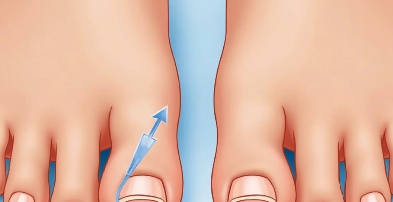
The space between your big toe and second toe might seem like a minor anatomical detail, but this gap holds significant clinical importance in podiatric medicine. This interdigital spacing, whether naturally occurring or developed over time, can reveal crucial information about foot biomechanics, underlying pathological conditions, and potential future complications. Understanding the implications of toe spacing variations helps both healthcare professionals and individuals recognise when this seemingly simple characteristic warrants closer attention.
From genetic predispositions to progressive deformities, the gap between the first and second toes serves as a window into the complex mechanics of foot function. This spacing can influence everything from gait patterns to shoe fitting, making it far more than a cosmetic consideration. Modern podiatric assessment now recognises this interdigital gap as a valuable diagnostic indicator that can guide treatment decisions and preventive care strategies.
Anatomical structure and terminology of interdigital spacing
The interdigital space between the big toe and second toe involves a complex arrangement of anatomical structures that work together to maintain proper foot alignment and function. This region encompasses soft tissue components, including the interdigital ligaments, plantar plate attachments, and intrinsic muscle insertions that collectively influence toe positioning and stability.
Hallux valgus interphalangeus and toe alignment mechanics
Hallux valgus interphalangeus represents a specific type of deviation where the big toe angles towards the second toe, creating or increasing the interdigital gap. This condition differs from traditional hallux valgus as the angulation occurs at the interphalangeal joint rather than the metatarsophalangeal joint. The mechanical forces that contribute to this alignment include imbalances in the flexor and extensor tendons, weakness in the intrinsic foot muscles, and alterations in the plantar aponeurosis tension.
The biomechanical implications of hallux valgus interphalangeus extend beyond simple toe positioning. When the big toe deviates laterally, it creates compensatory mechanisms throughout the forefoot that can lead to increased pressure on the second metatarsal head and potential stress fractures. This deviation also affects the windlass mechanism, a crucial component of normal gait that relies on proper big toe alignment for efficient propulsion.
Morton’s toe configuration and digital ray positioning
Morton’s toe, characterised by a second toe longer than the big toe, often coincides with increased interdigital spacing. This anatomical variation affects approximately 15-20% of the population and creates unique biomechanical challenges. The longer second toe bears additional weight during the stance phase of gait, potentially leading to increased pressure and compensatory toe spreading.
The relationship between Morton’s toe and interdigital gap formation involves complex interactions between the metatarsal lengths and the digital alignment. When the second metatarsal extends beyond the first, it creates an imbalance in weight distribution that can gradually increase the space between the first and second toes. This progression often develops slowly over years, making early recognition crucial for preventive intervention.
Metatarsophalangeal joint architecture and gap formation
The metatarsophalangeal joint architecture plays a fundamental role in determining interdigital spacing patterns. The joint capsule, collateral ligaments, and sesamoid complex all contribute to maintaining proper toe alignment. When these structures become compromised through injury, degenerative changes, or congenital variations, they can allow excessive toe deviation and gap formation.
Sesamoid positioning particularly influences the interdigital gap, as these small bones embedded within the flexor hallucis brevis tendon help maintain the big toe’s position relative to the first metatarsal. Sesamoid migration or fracture can disrupt this stabilising mechanism, leading to progressive toe deviation and increased spacing. Understanding these architectural relationships helps clinicians identify the underlying causes of gap formation and develop targeted treatment strategies.
Plantar plate integrity and interosseous muscle function
The plantar plate, a fibrocartilaginous structure that forms the floor of the metatarsophalangeal joint, provides crucial stability for toe positioning. Plantar plate tears or attenuation commonly occur in the second toe and can contribute to both hammer toe deformity and increased interdigital spacing. These injuries often result from repetitive stress or acute trauma and can be challenging to diagnose without advanced imaging.
Interosseous muscle dysfunction also plays a significant role in gap formation. The first dorsal interosseous muscle, responsible for abducting the second toe away from the big toe, can become overactive or hypertrophied in response to altered foot mechanics. Conversely, weakness in the adductor hallucis muscle can allow the big toe to drift away from the second toe, creating or exacerbating the interdigital gap.
Clinical assessment methods for interdigital gap measurement
Accurate assessment of interdigital spacing requires systematic evaluation using multiple measurement techniques and standardised protocols. Clinical assessment begins with visual inspection and manual examination but often requires advanced imaging and quantitative measurement tools to establish baseline values and monitor progression over time.
Radiographic evaluation using Weight-Bearing x-rays
Weight-bearing radiographs provide the gold standard for measuring interdigital spacing, as they capture the foot’s true functional position under load. Anteroposterior views allow for precise measurement of the intermetatarsal angle between the first and second metatarsals, which directly correlates with the clinical appearance of toe spacing. The normal intermetatarsal angle ranges from 8-10 degrees, with values exceeding 13 degrees indicating significant splaying.
Lateral radiographs complement the anteroposterior views by revealing sagittal plane deformities that may contribute to apparent toe spacing. Elevation of the first ray or depression of the second metatarsal head can create the illusion of increased gap without true horizontal separation. These radiographic findings help distinguish between structural and functional causes of interdigital spacing.
Podoscopic analysis and digital foot scanning techniques
Modern podoscopic analysis utilises pressure mapping technology to assess the functional implications of interdigital spacing during dynamic activities. These systems can identify areas of increased pressure concentration, altered load distribution patterns, and compensatory mechanisms that develop in response to toe spacing variations. The data collected helps quantify the clinical significance of measured gaps and guides treatment decisions.
Three-dimensional foot scanning technology has revolutionised the assessment of toe positioning and interdigital spacing. These systems capture precise measurements of toe angles, spacing dimensions, and volumetric changes over time. The non-invasive nature of digital scanning makes it ideal for monitoring progression and treatment response without radiation exposure.
Manchester scale grading system for hallux valgus
The Manchester Scale provides a standardised method for grading hallux valgus deformity severity, which directly relates to interdigital spacing patterns. This validated assessment tool uses photographic references to classify deformities into four grades, from normal alignment to severe deviation. The scale’s reliability and ease of use make it valuable for both clinical practice and research applications.
Incorporating Manchester Scale assessments into routine foot examinations helps track progression over time and communicate findings between healthcare providers. The visual nature of the scale also facilitates patient education by providing clear references for understanding their condition’s severity and potential for progression.
Intermetatarsal angle measurement protocols
Standardised protocols for measuring intermetatarsal angles ensure consistency and reproducibility in clinical assessments. The measurement technique involves identifying the longitudinal axis of the first and second metatarsals on weight-bearing radiographs and calculating the angle between these lines. Proper technique requires specific anatomical landmarks and standardised patient positioning to ensure accurate results.
Recent advances in digital radiographic measurement software have improved the precision and efficiency of angle calculations. These tools can automatically identify anatomical landmarks and calculate angles with greater accuracy than manual methods. However, understanding the underlying measurement principles remains crucial for interpreting results and identifying measurement errors.
Clinical photography documentation standards
Standardised clinical photography protocols ensure consistent documentation of interdigital spacing for monitoring and research purposes. Proper technique requires specific camera angles, lighting conditions, and patient positioning to capture accurate representations of toe alignment. The use of measurement scales or grid overlays helps quantify spacing dimensions from photographic images.
Digital photography integration with electronic health records facilitates longitudinal tracking of toe spacing changes and treatment outcomes. Comparative images taken at regular intervals provide valuable visual evidence of progression or improvement, supporting clinical decision-making and patient counselling regarding treatment options.
Pathological conditions associated with increased toe spacing
Various pathological conditions can contribute to abnormal interdigital spacing between the big toe and second toe. These conditions range from progressive deformities to inflammatory arthropathies, each presenting unique challenges for diagnosis and management. Understanding the underlying pathophysiology helps clinicians develop appropriate treatment strategies and predict long-term outcomes.
Progressive hallux valgus deformity stages
Hallux valgus deformity progresses through distinct stages, each characterised by increasing toe deviation and interdigital gap enlargement. The initial stage involves mild lateral deviation of the big toe with minimal gap formation, often accompanied by intermittent discomfort and minor shoe fitting difficulties. During this phase, conservative interventions may effectively halt or slow progression.
Moderate hallux valgus presents with more pronounced toe deviation and noticeable interdigital spacing. The increased gap often accompanies the development of a medial prominence (bunion) and may cause pressure on the second toe. Patients frequently report difficulty finding comfortable footwear and may experience pain during prolonged standing or walking activities.
Severe hallux valgus involves significant structural changes with marked interdigital spacing and potential second toe compression or override. The biomechanical alterations at this stage often require surgical intervention to restore proper alignment and function. Conservative management focuses primarily on symptom control and preventing further deterioration.
Bunion formation and medial eminence development
Bunion formation typically accompanies progressive interdigital gap enlargement as part of the hallux valgus deformity complex. The medial eminence develops through a combination of bony proliferation and soft tissue inflammation in response to altered biomechanical forces and external pressure from footwear. This prominence often becomes the primary source of symptoms, overshadowing the significance of the associated toe spacing.
The relationship between bunion severity and interdigital gap dimensions varies among individuals, influenced by factors such as bone quality, ligamentous laxity, and activity levels. Some patients develop large bunions with minimal toe spacing, while others exhibit significant gaps with relatively small medial prominences. This variability highlights the importance of comprehensive assessment rather than focusing solely on the most visible deformity component.
Crossover toe syndrome and digital instability
Crossover toe syndrome represents a progressive condition where the second toe gradually migrates over or under the big toe, often associated with increased interdigital spacing in the early stages. This condition typically begins with plantar plate attenuation or rupture, leading to progressive instability and eventual toe displacement. The initial presentation may include subtle increases in toe spacing before the characteristic crossing pattern develops.
Digital instability contributing to crossover toe syndrome can result from various factors, including trauma, inflammatory conditions, or biomechanical overload. Early recognition of increased interdigital spacing in at-risk patients can facilitate prompt intervention to prevent progression to frank crossover deformity. Conservative management during the early stages focuses on supporting the compromised plantar plate and maintaining proper toe alignment.
Rheumatoid arthritis impact on forefoot architecture
Rheumatoid arthritis profoundly affects forefoot architecture, often leading to characteristic patterns of toe spacing and deformity. The inflammatory process targets the synovial tissues within the metatarsophalangeal joints, leading to capsular distension, ligamentous laxity, and eventual joint destruction. These changes frequently result in hallux valgus deformity with associated interdigital gap formation.
The pattern of toe spacing in rheumatoid arthritis differs from mechanical hallux valgus in its rapid progression and bilateral symmetry. Patients often develop significant interdigital gaps within months rather than years, accompanied by subluxation or dislocation of the metatarsophalangeal joints. Management requires coordination between rheumatology and podiatric specialists to address both the underlying inflammatory process and the resulting structural deformities.
Genetic and developmental factors in toe gap formation
Genetic predisposition plays a substantial role in determining foot structure and the likelihood of developing interdigital spacing abnormalities. Family history studies demonstrate clear hereditary patterns in hallux valgus development, with maternal inheritance showing particularly strong correlations. Understanding these genetic factors helps identify at-risk individuals and implement preventive strategies before significant deformities develop.
Developmental factors during childhood and adolescence significantly influence adult foot structure and toe spacing patterns. Rapid growth periods, particularly during puberty, can unmask underlying structural predispositions and accelerate deformity progression. Early identification of risk factors, such as flexible flat feet or ligamentous laxity, allows for interventions during the formative years when structural modifications remain possible.
Congenital conditions affecting foot development can present with abnormal interdigital spacing from birth. Sandal gap deformity , characterised by increased spacing between the first and second toes, occurs in approximately 2-3% of newborns and may indicate underlying genetic syndromes or developmental abnormalities. While often benign as an isolated finding, this condition warrants careful evaluation to exclude associated anomalies.
Environmental factors during development, including footwear choices and activity patterns, interact with genetic predispositions to influence final toe spacing outcomes. Cultural practices regarding footwear, such as early introduction of restrictive shoes or prolonged barefoot activity, can modify the expression of genetic tendencies. Understanding these interactions helps clinicians provide culturally sensitive recommendations for preventing deformity progression.
Biomechanical implications for gait and weight distribution
The presence of increased interdigital spacing between the big toe and second toe creates significant biomechanical alterations that extend throughout the kinetic chain. These changes affect not only local foot function but can influence ankle, knee, hip, and even spinal alignment during walking and running activities. The big toe’s role in the terminal stance phase of gait becomes compromised when it deviates laterally, reducing the efficiency of the push-off mechanism.
Weight distribution patterns change substantially when interdigital spacing increases beyond normal parameters. The first ray, comprising the big toe and first metatarsal, typically bears approximately 40-60% of the forefoot loading during normal gait. When the big toe deviates laterally and creates increased spacing, this load transfers to the central metatarsals, particularly the second and third rays. This redistribution can lead to metatarsalgia, stress fractures, and callus formation under the lesser metatarsal heads.
Compensatory mechanisms develop throughout the lower extremity to accommodate the altered foot mechanics associated with increased toe spacing. The subtalar joint may increase pronation to maintain ground contact with the medially deviated first ray, while the ankle complex adjusts to preserve overall stability. These compensations, while initially protective, can lead to overuse injuries in the ankle, Achilles tendon, and posterior tibial tendon over time.
Dynamic foot function during athletic activities becomes particularly challenging when significant interdigital spacing exists. Sports requiring rapid direction changes, jumping, or sustained running place increased demands on the forefoot’s stability mechanisms. Athletes with increased toe spacing may experience reduced performance due to decreased push-off efficiency and increased energy expenditure during propulsion phases. Understanding these biomechanical implications helps guide activity modifications and performance optimisation strategies.
The relationship between interdigital spacing and overall postural control demonstrates the foot’s crucial role in maintaining balance and stability. Proprioceptive feedback from the toe spacing region contributes to the central nervous system’s understanding of foot position and ground contact. Alterations in this feedback mechanism can affect balance reactions and increase fall risk, particularly in elderly individuals or those with concurrent neurological conditions.
Treatment approaches and corrective interventions
Conservative treatment strategies form the foundation of management for most cases of increased interdigital spacing, particularly in the early stages of deformity development. These approaches focus on addressing the underlying biomechanical factors contributing to toe deviation while providing symptom relief and preventing progression. The success of conservative interventions depends largely on early recognition and patient compliance with recommended modifications.
Orthotic interventions represent a cornerstone of conservative management, with custom-designed devices offering the most precise correction for individual foot mechanics. Metatarsal pads , when properly positioned, can redistribute plantar pressures and encourage more normal toe alignment during weight-bearing activities. The positioning of these pads requires expertise, as incorrect placement can exacerbate existing problems or create new areas of pressure concentration.
Footwear modifications play a crucial role in managing symptoms and preventing progression of interdigital spacing abnormalities. Shoes with adequate toe box width and depth accommodate the altered toe
positioning without creating additional pressure points. Wide toe box designs allow the toes to maintain their natural spacing without compression, while adequate depth prevents dorsal pressure on hammer toes that commonly develop alongside increased interdigital spacing.
Exercise therapy targeting the intrinsic foot muscles can help improve toe alignment and reduce progression of spacing abnormalities. Specific exercises focusing on toe flexion, extension, and abduction help strengthen the muscles responsible for maintaining proper digital alignment. Towel scrunches, marble pickups, and toe spreading exercises performed daily can provide measurable improvements in muscle strength and coordination over time.
Taping techniques offer temporary but effective correction of toe positioning during activities that place increased stress on the forefoot. Buddy taping the big toe to the second toe can provide stability and reduce symptoms during sports participation or prolonged walking. However, these techniques require proper instruction to avoid circulatory compromise or skin irritation from prolonged application.
Surgical interventions become necessary when conservative measures fail to provide adequate symptom relief or when deformity progression threatens foot function. The choice of surgical procedure depends on the underlying cause of the interdigital spacing, the degree of deformity, and the patient’s functional requirements. Early intervention typically involves soft tissue procedures, while advanced deformities may require bony corrections or joint fusions.
Minimally invasive surgical techniques have revolutionised the treatment of hallux valgus and associated interdigital spacing abnormalities. Percutaneous osteotomies and soft tissue releases can be performed through small incisions, reducing surgical trauma and accelerating recovery times. These techniques require specialised training and equipment but offer significant advantages in terms of cosmetic outcomes and return to activities.
Bunionectomy procedures address both the medial prominence and the underlying toe deviation responsible for increased interdigital spacing. Modern surgical techniques focus on correcting the first ray alignment while preserving joint motion and maintaining the foot’s natural biomechanics. The choice between different bunionectomy procedures depends on factors such as the severity of deformity, bone quality, and patient age.
Post-surgical rehabilitation plays a crucial role in achieving optimal outcomes and preventing recurrence of toe spacing abnormalities. Early mobilisation protocols help maintain joint flexibility while protecting the surgical correction during the healing process. Progressive weight-bearing and return to activities must be carefully supervised to ensure proper healing and long-term stability of the correction.
Emerging treatment modalities, including regenerative medicine techniques and advanced biomaterial implants, show promise for addressing the underlying tissue defects that contribute to interdigital spacing abnormalities. Platelet-rich plasma injections may help strengthen compromised plantar plate structures, while synthetic ligament replacements could provide long-term stability for severely damaged joints. These innovative approaches require further research but represent exciting possibilities for future treatment options.
Patient education remains a critical component of any treatment plan for interdigital spacing abnormalities. Understanding the progressive nature of these conditions motivates patients to comply with conservative interventions and seek timely surgical consultation when indicated. Regular monitoring and adjustment of treatment strategies help optimise outcomes and prevent complications that could compromise long-term foot function.
The integration of technology into treatment planning has enhanced precision and predictability of interventions for toe spacing abnormalities. Computer-assisted surgical planning allows surgeons to simulate corrections and predict outcomes before performing procedures. Similarly, 3D printing technology enables the creation of custom orthotic devices and surgical guides that improve the accuracy of interventions.
Long-term follow-up studies demonstrate the importance of ongoing monitoring even after successful treatment of interdigital spacing abnormalities. Recurrence rates vary depending on the treatment method and underlying factors, with some patients requiring additional interventions over time. Understanding these patterns helps clinicians counsel patients appropriately and develop realistic expectations for treatment outcomes.