Complement proteins C3 and C4 serve as critical components of the immune system’s defence mechanisms, functioning as molecular sentries that detect and neutralise potential threats to human health. When laboratory tests reveal low C3 and C4 levels , this finding often signals underlying immune system dysfunction or active disease processes that warrant immediate clinical attention. These complement proteins operate within a sophisticated cascade system, where reduced concentrations can indicate everything from autoimmune conditions like systemic lupus erythematosus to infectious diseases and inherited immunodeficiencies.
Understanding the clinical significance of decreased complement levels requires appreciation of their complex roles in immune surveillance, pathogen elimination, and tissue homeostasis. Healthcare professionals routinely measure these proteins to diagnose conditions ranging from glomerulonephritis to hereditary angioedema, making their interpretation essential for effective patient management. The intricate relationship between complement consumption, synthesis, and disease activity creates a dynamic biomarker system that reflects real-time immune system status.
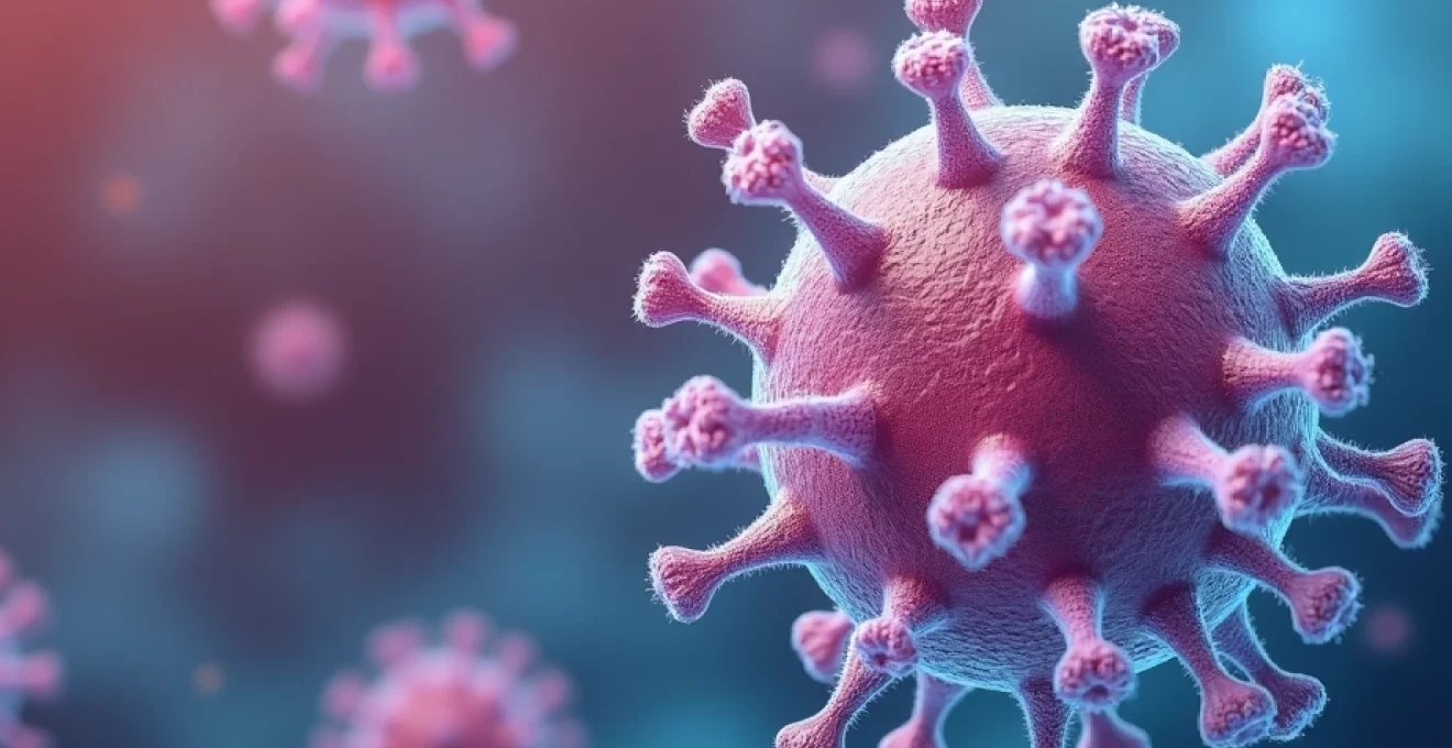
Complement C3 and C4 proteins: molecular structure and function in immune defence
The complement system represents one of the most sophisticated molecular defence networks in human biology, comprising over 30 distinct proteins that work in coordinated sequence to protect against microbial invasion and maintain tissue integrity. C3 and C4 proteins function as central mediators within this cascade, each possessing unique structural characteristics that enable their specific immunological roles. C3, weighing approximately 185 kilodaltons, exists as the most abundant complement protein in human serum, with normal concentrations ranging from 0.65 to 1.90 grams per litre depending on the laboratory reference standards employed.
C4 protein, though present in lower concentrations typically between 0.14 and 0.54 grams per litre, plays an equally crucial role in immune recognition and activation. The molecular architecture of these proteins includes reactive thioester bonds that become exposed upon activation, allowing covalent attachment to target surfaces such as bacterial cell walls or immune complexes. This attachment mechanism transforms these proteins from circulating sentries into surface-bound markers that facilitate subsequent immune responses including phagocytosis and membrane attack complex formation.
C3 convertase formation and alternative pathway activation
The formation of C3 convertase represents a pivotal amplification point in complement activation, where small initiating signals become magnified into robust immune responses. Alternative pathway C3 convertase, designated as C3bBb , forms through the spontaneous hydrolysis of C3 and subsequent binding of factor B, which becomes cleaved by factor D to generate the catalytically active Bb fragment. This convertase complex exhibits remarkable efficiency, capable of cleaving hundreds of C3 molecules per minute when properly regulated by complement control proteins.
Factor H and factor I serve as critical regulators of alternative pathway activation, preventing excessive complement consumption on healthy tissue surfaces while permitting activation on pathogenic targets. When these regulatory mechanisms become compromised through genetic mutations or acquired deficiencies, uncontrolled C3 consumption can occur, leading to persistently low serum levels and increased susceptibility to infections.
C4 classical pathway integration with immunoglobulin complexes
Classical pathway activation begins when C1q recognises and binds to immune complexes containing IgG or IgM antibodies, initiating a conformational change that activates the associated C1r and C1s proteases. Activated C1s cleaves C4 into C4a and C4b fragments, with C4b depositing covalently onto nearby surfaces through its exposed thioester bond. This C4b deposition creates binding sites for C2, which becomes cleaved by C1s to form the classical pathway C3 convertase C4b2a .
The classical pathway demonstrates exquisite specificity for antibody-antigen complexes, making C4 consumption a sensitive marker for immune complex-mediated diseases. Conditions such as systemic lupus erythematosus frequently exhibit low C4 levels due to ongoing classical pathway activation by circulating immune complexes, creating a valuable diagnostic and monitoring parameter for disease activity assessment.
Complement cascade amplification through c3b opsonisation
C3b molecules generated through convertase activity serve dual functions as opsonins that enhance phagocytosis and as building blocks for further complement activation. Deposited C3b molecules create recognition signals for complement receptors on phagocytic cells, dramatically increasing the efficiency of pathogen clearance. Additionally, C3b can bind additional factor B molecules to form new alternative pathway convertases, creating positive feedback loops that amplify the initial activation signal.
This amplification mechanism explains why relatively modest complement activation can lead to substantial C3 consumption and correspondingly low serum levels. The balance between complement activation and regulation determines whether this amplification serves protective functions or contributes to tissue damage through excessive inflammatory responses.
Terminal complement complex assembly via C5 convertase
C5 convertases form when C3b associates with existing C3 convertases, creating enzymatic complexes capable of cleaving C5 into C5a and C5b fragments. C5b serves as the nucleation site for terminal complement complex assembly, sequentially binding C6, C7, C8, and multiple C9 molecules to form the membrane attack complex (MAC). This MAC structure creates pores in target cell membranes, leading to osmotic lysis and pathogen destruction.
Terminal pathway activation requires adequate C3 and C4 levels for efficient convertase formation, explaining why deficiencies in these proteins can compromise overall complement function despite normal levels of terminal components. The measurement of CH50, which assesses the functional capacity of the entire classical pathway, often reveals reduced activity when C3 or C4 levels are significantly decreased.
Clinical manifestations associated with reduced C3 and C4 complement levels
Low complement levels manifest through diverse clinical presentations that reflect the multifaceted roles of these proteins in immune surveillance and inflammatory regulation. Patients with decreased C3 and C4 concentrations frequently present with increased susceptibility to bacterial infections, particularly those caused by encapsulated organisms such as Streptococcus pneumoniae and Neisseria species. This enhanced infection risk stems from impaired opsonisation and reduced bactericidal activity, creating clinical scenarios where routine infections become severe or recurrent.
Autoimmune manifestations represent another major category of clinical presentations associated with complement deficiency states . Paradoxically, while complement activation can contribute to autoimmune tissue damage, complement deficiencies often predispose to autoimmune disease development through impaired clearance of immune complexes and apoptotic cells. This dual role creates complex clinical scenarios where complement measurement becomes essential for both diagnosis and therapeutic monitoring.
Systemic lupus erythematosus and immune complex deposition
Systemic lupus erythematosus exemplifies the complex relationship between complement levels and autoimmune disease activity. During active disease phases, immune complex formation and deposition lead to classical pathway activation and subsequent consumption of C3 and C4 proteins. Laboratory studies consistently demonstrate inverse correlations between disease activity indices and complement levels, with the most severe lupus flares typically associated with profound complement depletion.
Lupus nephritis represents a particularly concerning manifestation where low complement levels correlate with renal immune complex deposition and glomerular inflammation. Serial monitoring of C3 and C4 levels enables clinicians to assess treatment responses and predict renal flare episodes, making these measurements indispensable tools in lupus management strategies.
Hereditary angioedema type II C1 esterase inhibitor deficiency
Hereditary angioedema type II presents a unique complement abnormality where C1 esterase inhibitor dysfunction leads to episodic complement activation and C4 consumption. Unlike lupus-associated complement depletion, hereditary angioedema typically demonstrates isolated C4 depression with normal C3 levels, reflecting specific classical pathway activation without alternative pathway involvement. Episodes of angioedema coincide with periods of increased complement consumption, creating diagnostic opportunities through complement measurement during acute presentations.
The clinical presentation includes recurrent episodes of non-pitting oedema affecting the face, extremities, and potentially life-threatening laryngeal involvement. C4 levels often remain chronically depressed in affected individuals, serving as a screening tool for this potentially fatal condition that requires specific therapeutic interventions including C1 esterase inhibitor replacement therapy.
Membranoproliferative glomerulonephritis and renal complement consumption
Membranoproliferative glomerulonephritis encompasses several distinct pathological entities characterised by glomerular complement deposition and consumption. Type II disease, also known as dense deposit disease, frequently presents with persistently low C3 levels due to alternative pathway dysregulation caused by C3 nephritic factors or complement regulatory protein deficiencies. These autoantibodies stabilise C3 convertases, leading to continuous alternative pathway activation and C3 consumption.
Patients typically present with haematuria, proteinuria, and progressive renal dysfunction accompanied by characteristically low C3 levels that may persist for months or years. The combination of renal manifestations with prolonged complement depletion creates a distinctive clinical pattern that aids in differential diagnosis from other forms of glomerulonephritis where complement levels normalise more rapidly following treatment.
Acquired complement deficiencies in chronic liver disease
Chronic liver disease represents an often-overlooked cause of complement deficiency through impaired hepatic synthesis of complement proteins. The liver produces the majority of circulating complement components, making hepatic dysfunction a significant contributor to low C3 and C4 levels. Unlike consumption-based complement depletion, liver disease typically produces proportional decreases in multiple complement components corresponding to the severity of hepatic synthetic dysfunction.
Patients with cirrhosis or severe hepatitis may demonstrate complement levels that correlate with other markers of hepatic synthetic function, including albumin and transferrin concentrations. This pattern of complement deficiency contributes to the increased infection susceptibility observed in patients with advanced liver disease, particularly spontaneous bacterial peritonitis and other serious bacterial complications.
Laboratory diagnostic methods for C3 and C4 quantification
Modern complement measurement relies on sophisticated analytical techniques that provide both quantitative protein concentrations and functional activity assessments. The choice of analytical method significantly influences result interpretation, with each technique offering distinct advantages and limitations that affect clinical decision-making. Immunochemical methods measure protein concentrations regardless of functional activity, while haemolytic assays assess the biological activity of complement pathways under standardised conditions.
Quality control measures become particularly critical in complement analysis due to the labile nature of these proteins and their susceptibility to degradation during sample collection and processing. Proper specimen handling, including rapid separation of serum and appropriate storage conditions, directly impacts result accuracy and clinical relevance. Many laboratories now employ multiple analytical approaches to provide comprehensive complement assessment that addresses both quantitative and functional parameters.
Radial immunodiffusion techniques using mancini plates
Radial immunodiffusion represents one of the earliest quantitative methods for complement protein measurement, utilising antibody-containing agarose gels to create precipitation rings proportional to antigen concentration. This technique offers excellent precision for C3 and C4 quantification, with results typically available within 24-48 hours of sample processing. The method’s reliance on diffusion kinetics provides inherent quality control through ring formation patterns that reveal sample integrity issues or technical problems.
Despite the availability of more rapid analytical methods, radial immunodiffusion maintains clinical relevance for reference standard preparation and method validation. The technique’s ability to detect both native and activated complement proteins makes it particularly valuable for research applications investigating complement activation patterns in various disease states.
Nephelometry and turbidimetric immunoassays for rapid analysis
Nephelometric and turbidimetric immunoassays have become the standard methods for routine complement quantification in clinical laboratories worldwide. These techniques measure light scatter or absorption changes that occur when specific antibodies form immune complexes with complement proteins, providing results within minutes of sample introduction. Automated analysers enable high-throughput processing with excellent precision and minimal operator intervention, making these methods ideal for busy clinical laboratories.
The primary advantages include rapid turnaround times, typically 2-3 days for most laboratories, and the ability to process multiple samples simultaneously. However, these methods measure total protein concentration rather than functional activity, potentially missing functionally deficient but antigenically normal proteins that could affect clinical interpretation.
CH50 haemolytic complement activity functional testing
CH50 testing represents the gold standard for assessing classical complement pathway function, measuring the serum dilution required to lyse 50% of antibody-sensitised sheep erythrocytes under standardised conditions. This functional assay integrates the activity of all classical pathway components, providing a sensitive indicator of complement system integrity that correlates closely with in vivo biological activity. Results typically range from 150-300 CH50 units per millilitre in healthy individuals, with complete pathway deficiencies producing undetectable activity.
The assay’s ability to detect functionally significant abnormalities makes it invaluable for evaluating patients with recurrent infections or suspected complement deficiencies. However, the requirement for fresh complement components and standardised red blood cell preparations creates logistical challenges that limit availability in some laboratory settings.
AH50 alternative pathway assessment protocols
Alternative pathway assessment through AH50 testing employs rabbit erythrocytes in EGTA-magnesium buffer to selectively activate the alternative pathway while chelating calcium required for classical pathway function. This specialised functional assay becomes particularly valuable when alternative pathway-specific abnormalities are suspected, such as those seen in certain forms of glomerulonephritis or complement regulatory protein deficiencies.
The combination of CH50 and AH50 testing enables differentiation between classical and alternative pathway defects, guiding further diagnostic evaluation and therapeutic planning. Normal CH50 with reduced AH50 suggests alternative pathway-specific abnormalities, while the converse pattern indicates classical pathway dysfunction with preserved alternative pathway function.
Therapeutic interventions for complement deficiency management
Treatment strategies for complement deficiency states vary dramatically depending on the underlying aetiology, ranging from immunosuppressive therapy for autoimmune-mediated consumption to protein replacement therapy for hereditary deficiencies. The selection of appropriate therapeutic interventions requires careful consideration of disease mechanisms, complement pathway specificity, and individual patient factors including infection risk and treatment tolerance. Modern complement therapeutics increasingly target specific pathway components, enabling more precise interventions with reduced systemic effects.
Emerging therapeutic approaches include complement inhibitors such as eculizumab for terminal pathway blockade and C1 esterase inhibitor concentrates for hereditary angioedema management. These targeted therapies represent significant advances over traditional broad-spectrum immunosuppression, offering improved efficacy with fewer adverse effects. The development of novel complement modulators continues to expand therapeutic options for patients with previously intractable complement-related disorders.
Supportive care measures play equally important roles in complement deficiency management, particularly regarding infection prevention and early recognition of complications. Vaccination strategies become critical considerations, with enhanced immunisation protocols often recommended for patients with significant complement defects. However, live vaccines may be contraindicated in certain complement deficiency states, requiring individualised risk-benefit assessments for optimal patient care.
The integration of complement testing with clinical presentation creates powerful diagnostic tools that guide therapeutic decision-making and enable personalised treatment approaches for complex immune system disorders.
Differential diagnosis: distinguishing primary from secondary complement depletion
The differential diagnosis of low complement levels requires systematic evaluation of potential causes, ranging from inherited complement component deficiencies to acquired consumption states associated with active disease processes. Primary complement deficiencies, though rare, typically present with characteristic patterns of complement abnormalities that persist despite treatment of associated conditions. These genetic defects affect individual complement components or regulatory proteins, creating specific vulnerability patterns that guide diagnostic evaluation and therapeutic planning.
Secondary complement consumption represents a more common clinical scenario where active disease processes lead to complement activation and depletion through increased utilisation rather than reduced synthesis. Distinguishing between these mechanisms becomes critical for treatment selection, as primary deficiencies may require protein replacement therapy while secondary depletion typically responds to treatment of the underlying disease process. The temporal relationship between complement levels and disease activity provides valuable diagnostic clues, with secondary depletion typically demonstrating improvement following successful treatment of the precipitating condition.
Laboratory evaluation strategies must consider the specific patterns of complement abnormalities observed in different clinical scenarios. Isolated C4 deficiency with normal C3 levels suggests classical pathway-specific problems such as hereditary angioedema or early-stage lupus, while combined C3 and C4 depletion typically indicates more extensive complement activation or synthesis problems. The integration of functional testing with quantitative measurements provides additional discrimination between these diagnostic possibilities,
enhancing clinical diagnostic accuracy and therapeutic monitoring capabilities.
Molecular genetic analysis has revolutionised the identification of primary complement deficiencies, enabling precise characterisation of mutations affecting complement component synthesis or function. These hereditary conditions typically demonstrate consistent complement abnormalities that persist regardless of disease activity, contrasting sharply with the fluctuating levels observed in secondary consumption states. Family history evaluation becomes crucial in suspected primary deficiencies, as these conditions often follow predictable inheritance patterns that support diagnostic hypotheses.
The clinical presentation timeline provides additional diagnostic insights, with primary deficiencies typically manifesting early in life through recurrent infections or unusual disease susceptibility patterns. Secondary complement depletion usually develops in association with recognisable disease processes, creating temporal relationships that support diagnostic differentiation. Age of onset, family history, and response to therapeutic interventions collectively contribute to accurate diagnostic classification and appropriate treatment selection.
Prognostic implications and long-term monitoring strategies for hypocomplementaemia
Long-term prognosis in patients with persistently low complement levels depends heavily on the underlying aetiology and response to therapeutic interventions. Patients with autoimmune-mediated complement consumption generally demonstrate favourable outcomes when disease activity is successfully controlled through immunosuppressive therapy, with complement levels serving as valuable biomarkers for treatment response assessment. However, those with primary complement deficiencies face lifelong increased infection susceptibility that requires ongoing vigilance and preventive strategies to minimise complications.
Monitoring strategies must be tailored to individual patient circumstances, with frequency and intensity of surveillance adjusted based on disease activity, treatment response, and clinical stability. Serial complement measurements provide early warning signs of disease flares in conditions like systemic lupus erythematosus, enabling preemptive therapeutic adjustments that can prevent serious complications such as lupus nephritis or central nervous system involvement. The integration of complement monitoring with other disease-specific biomarkers creates comprehensive assessment frameworks that optimise long-term patient outcomes.
Risk stratification becomes particularly important for patients with significant complement deficiencies, as their increased susceptibility to serious infections requires proactive management approaches. Vaccination strategies, antibiotic prophylaxis considerations, and early intervention protocols must be carefully planned to maximise protection while minimising unnecessary interventions. Regular assessment of infection patterns, treatment responses, and quality of life measures enables ongoing refinement of management strategies to meet evolving patient needs.
The development of personalised monitoring protocols requires consideration of multiple factors including disease type, complement deficiency pattern, treatment regimen, and individual risk factors. Patients with classical pathway defects may require different surveillance approaches compared to those with alternative pathway abnormalities, reflecting the distinct clinical manifestations associated with each pathway dysfunction. The emergence of novel therapeutic agents continues to influence monitoring strategies, as new treatments may require specific biomarker assessments to optimise efficacy and minimise adverse effects.
Successful long-term management of complement deficiency states requires multidisciplinary collaboration between immunologists, nephrologists, rheumatologists, and infectious disease specialists to address the complex interplay between immune dysfunction, infection risk, and autoimmune manifestations.
Quality of life considerations become increasingly important as patients with chronic complement deficiencies navigate long-term management challenges. The psychological impact of chronic illness, treatment burden, and infection anxiety requires ongoing attention and support to maintain optimal patient wellbeing. Educational initiatives that empower patients to recognise early warning signs of complications and understand their condition facilitate improved self-management and treatment adherence.
Future developments in complement research promise continued improvements in diagnostic accuracy and therapeutic options for patients with complement deficiency states. Advances in molecular diagnostics, novel therapeutic targets, and personalised medicine approaches will likely transform the management landscape for these complex conditions. The integration of artificial intelligence and machine learning technologies may enable more sophisticated prediction models for disease progression and treatment response, further enhancing patient outcomes through precision medicine approaches.
The establishment of patient registries and collaborative research networks continues to expand our understanding of complement deficiency states and their long-term implications. These initiatives provide valuable insights into natural disease progression, treatment effectiveness, and optimal management strategies that benefit the broader medical community and improve care for affected patients. As our knowledge base continues to evolve, the prognosis for patients with complement deficiencies will likely continue to improve through enhanced diagnostic capabilities and targeted therapeutic interventions.