Understanding the precise dimensions and characteristics of normal thyroid lobes forms the cornerstone of accurate endocrine assessment and clinical decision-making. The thyroid gland, a butterfly-shaped endocrine organ positioned in the anterior neck, exhibits specific dimensional parameters that serve as critical benchmarks for identifying pathological conditions. These measurements have become increasingly important as ultrasonography continues to evolve as the primary imaging modality for thyroid evaluation. Healthcare professionals rely on established reference values to distinguish between physiological variations and pathological enlargement, making comprehensive knowledge of normal thyroid dimensions essential for optimal patient care.
Anatomical dimensions and morphological characteristics of healthy thyroid lobes
The thyroid gland demonstrates remarkable consistency in its structural dimensions across healthy populations, with each lobe exhibiting specific morphological characteristics that define normalcy. Understanding these parameters enables clinicians to establish baseline measurements and identify deviations that may indicate underlying pathology.
Standard anteroposterior, transverse, and craniocaudal measurements
Normal thyroid lobe dimensions follow well-established patterns that vary according to age and developmental stage. In newborns, each lobe typically measures 18-20 mm in the longitudinal axis and 8-9 mm in the anteroposterior diameter. These dimensions reflect the gland’s initial developmental state and provide essential reference points for paediatric thyroid assessment.
By one year of age, significant growth occurs, with longitudinal measurements reaching approximately 25 mm and anteroposterior diameters expanding to 12-15 mm. This rapid expansion during the first year of life corresponds to the increasing metabolic demands of the developing infant and the maturation of thyroid hormone synthesis pathways.
Adult thyroid lobes demonstrate more stable dimensions, typically measuring 40-60 mm in the longitudinal axis and 13-18 mm in the anteroposterior diameter. These measurements represent the fully developed gland and serve as the primary reference standards for clinical assessment. The transverse dimension usually ranges from 12-20 mm, completing the three-dimensional profile necessary for accurate volumetric calculations.
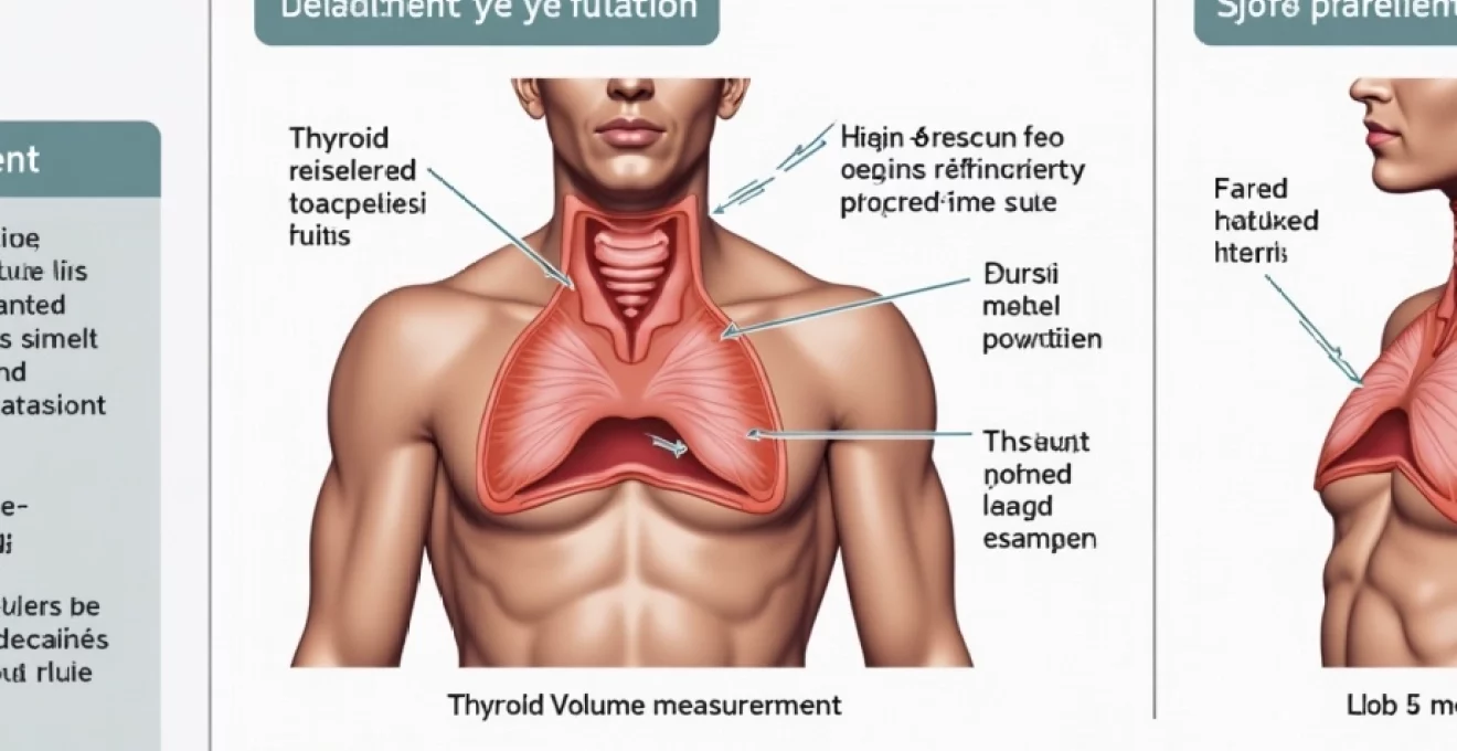
Thyroid volume calculations using ellipsoid formula methods
Precise thyroid volume determination relies on the ellipsoid formula, which accounts for the gland’s characteristic three-dimensional shape. This mathematical approach provides more accurate assessments than simple dimensional measurements alone. The standard ellipsoid formula calculates volume by multiplying length × width × depth × 0.523, where 0.523 represents the correction factor for ellipsoid geometry.
Normal thyroid volume ranges demonstrate gender-specific variations that reflect physiological differences between males and females. For healthy adult women, total thyroid volume typically falls within 10-15 ml, whilst men generally exhibit slightly larger volumes ranging from 12-18 ml. These differences correlate with body size variations and hormonal influences on thyroid growth patterns.
When calculating individual lobe volumes, healthcare professionals must consider both lobes separately before determining total gland volume. Each lobe contributes approximately equally to the total volume, though minor asymmetries are common and considered normal. The isthmus volume should be included in total calculations when its thickness exceeds 3 mm, as this additional tissue contributes meaningfully to overall thyroid mass.
Right versus left lobe asymmetry patterns in normal adults
Physiological asymmetry between thyroid lobes represents a normal finding that occurs in most healthy individuals. Research demonstrates that the right lobe frequently measures slightly larger than the left lobe, with differences of up to 15-20% considered within normal limits. This asymmetry likely reflects developmental variations and vascular supply differences between the two sides.
The degree of acceptable asymmetry varies across populations and measurement techniques. Ultrasonographic studies consistently show that complete symmetry between lobes is actually uncommon, with most individuals demonstrating some degree of size variation. Clinical significance emerges only when asymmetry exceeds 25% or when one lobe appears structurally abnormal compared to its counterpart.
Understanding normal asymmetry patterns prevents unnecessary alarm during routine examinations and helps distinguish physiological variations from pathological changes. Significant asymmetry warranting further investigation typically involves dramatic size differences, structural abnormalities, or changes in echogenic characteristics that suggest underlying disease processes.
Age-related changes in thyroid lobe dimensions
Thyroid dimensions demonstrate predictable changes throughout the human lifespan, reflecting evolving metabolic requirements and hormonal influences. During infancy and childhood, rapid growth occurs as the gland develops its full functional capacity. Adolescence brings additional dimensional changes, particularly in girls, where hormonal fluctuations can cause temporary enlargement that typically resolves after puberty.
Adult thyroid dimensions remain relatively stable throughout most of middle age, though subtle changes may occur in response to iodine intake variations, pregnancy, or other physiological stressors. Elderly individuals often experience slight reductions in thyroid size, correlating with decreased metabolic demands and age-related tissue changes.
Pregnancy represents a special consideration, as physiological enlargement commonly occurs during gestation. This temporary expansion reflects increased metabolic demands and hormonal stimulation, typically resolving within months after delivery. Understanding these pregnancy-related changes prevents misinterpretation of normal physiological adaptations as pathological conditions.
Ultrasonographic assessment protocols for thyroid lobe evaluation
Modern thyroid assessment relies heavily on ultrasonographic imaging, which provides detailed visualisation of thyroid structure, dimensions, and internal characteristics. Proper technique and standardised protocols ensure consistent, reliable measurements that support accurate clinical decision-making.
High-frequency linear transducer positioning techniques
Optimal thyroid imaging requires high-frequency linear transducers, typically operating at 10-15 MHz, which provide excellent resolution for superficial structures. Patient positioning plays a crucial role in obtaining accurate measurements, with the neck placed in hyperextension to maximise gland exposure and improve visualisation of all thyroid regions.
Transducer manipulation follows the PART methodology: Pressure, Alignment, Rotation, and Tilt. Appropriate pressure ensures good skin contact whilst avoiding excessive compression that might distort gland dimensions. Proper alignment positions the target structure optimally within the scanning plane, whilst rotation and tilt adjustments optimise the angle of incidence for maximum reflection.
Both longitudinal and transverse scanning planes are essential for comprehensive assessment. Longitudinal views provide measurements of the craniocaudal and anteroposterior dimensions, whilst transverse views capture the mediolateral measurements necessary for volume calculations. Systematic scanning of each lobe ensures complete evaluation and reduces the likelihood of missing significant abnormalities.
Doppler flow assessment and vascular pattern analysis
Colour Doppler imaging provides valuable information about thyroid vascularity, which can indicate normal versus abnormal physiological states. Normal thyroid tissue demonstrates moderate vascularity with vessels distributed throughout the gland parenchyma. The BART mnemonic (Blue Away, Red Toward) helps interpret flow direction relative to the transducer position.
Healthy thyroid lobes typically show uniform vascular distribution without areas of significantly increased or decreased perfusion. Hypervascular patterns, sometimes described as thyroid inferno , may indicate conditions such as Graves’ disease or acute thyroiditis. Conversely, areas of decreased vascularity might suggest fibrosis, chronic inflammation, or other pathological processes.
Doppler assessment also evaluates the major vessels supplying the thyroid, including the superior and inferior thyroid arteries. Normal flow patterns in these vessels provide additional confirmation of physiological function and help identify vascular abnormalities that might affect gland function or surgical planning.
Echogenicity grading systems and texture classification
Normal thyroid tissue demonstrates characteristic ultrasonographic features that serve as reference standards for identifying abnormalities. Healthy thyroid parenchyma appears homogeneously hyperechoic compared to adjacent neck muscles, reflecting its high iodine content. This increased echogenicity creates the classic ground glass appearance that experienced ultrasonographers recognise immediately.
Echo texture classification systems help standardise reporting and improve communication between healthcare providers. Fine, uniform echotexture represents normal thyroid tissue, whilst coarse or heterogeneous patterns may indicate pathological changes. Small echo-free zones measuring 1-2 mm typically represent normal blood vessels and should not be mistaken for pathological cysts or nodules.
Normal thyroid tissue exhibits a homogeneous, hyperechoic appearance with fine internal texture that reflects optimal iodine content and healthy cellular architecture.
Measurement standardisation according to AACE guidelines
The American Association of Clinical Endocrinologists (AACE) has established comprehensive guidelines for thyroid ultrasonography that promote consistency and accuracy in clinical practice. These protocols specify measurement techniques, reporting standards, and quality assurance measures that ensure reliable results across different practitioners and institutions.
Standardised measurement protocols require three-dimensional assessment of each thyroid lobe, with measurements taken at the maximal dimensions in each plane. Length measurements are obtained in the longitudinal plane, width measurements in the transverse plane, and depth measurements represent the anteroposterior dimension. The isthmus thickness should be measured in the transverse plane and documented separately.
Documentation requirements include permanent image storage with appropriate patient identification, facility marking, examination date, and anatomical orientation labels. Quality assurance measures emphasise equipment calibration, technique consistency, and regular competency assessment for personnel performing thyroid examinations.
Reference values and Population-Based normative data
Establishing accurate reference ranges requires extensive population studies that account for demographic, geographic, and physiological variables. These normative databases provide the foundation for clinical decision-making and help distinguish normal variations from pathological conditions requiring intervention.
Gender-specific thyroid lobe size variations in european populations
European population studies reveal consistent patterns of gender-related thyroid size differences across diverse ethnic and geographic groups. Men typically demonstrate 15-20% larger thyroid volumes compared to women, reflecting differences in body size, metabolic rate, and hormonal influences. These variations persist across age groups and appear consistent regardless of iodine intake status.
Regional variations within Europe suggest that genetic factors, dietary patterns, and environmental influences may contribute to population-specific reference ranges. Nordic countries often report slightly smaller average thyroid volumes compared to Mediterranean regions, possibly reflecting differences in iodine availability and dietary habits throughout historical periods.
Age-stratified reference ranges provide more precise guidance for clinical interpretation, as thyroid dimensions change predictably throughout life. Paediatric reference ranges require separate consideration due to rapid growth patterns during childhood and adolescence, whilst elderly populations may demonstrate different normal ranges reflecting age-related physiological changes.
Body mass index correlation with thyroid volume parameters
Research consistently demonstrates positive correlations between body mass index (BMI) and thyroid volume, suggesting that larger individuals typically have proportionally larger thyroid glands. This relationship appears linear across most BMI ranges and provides important context for interpreting thyroid measurements in clinical practice.
The correlation between BMI and thyroid volume likely reflects increased metabolic demands associated with larger body mass, requiring greater thyroid hormone production capacity. However, the relationship is not simple linear scaling, as other factors such as insulin resistance, inflammatory markers, and metabolic syndrome components may influence thyroid growth patterns independently.
Clinical implications of BMI-thyroid volume relationships suggest that reference ranges should ideally be adjusted for body size to improve diagnostic accuracy. Some practitioners use body surface area calculations to normalise thyroid volume measurements, whilst others rely on BMI-stratified reference ranges to guide clinical interpretation.
Iodine sufficiency impact on normal thyroid dimensions
Iodine availability significantly influences normal thyroid dimensions, with populations experiencing chronic iodine deficiency typically demonstrating larger average thyroid volumes. This compensatory enlargement represents the gland’s attempt to maximise iodine capture and maintain adequate hormone production despite limited substrate availability.
Geographic regions with optimal iodine intake, typically achieved through iodised salt programs, demonstrate smaller average thyroid volumes and more consistent population reference ranges. The transition from iodine deficiency to sufficiency in various populations has provided valuable insights into the relationship between iodine status and thyroid morphology.
Adequate iodine intake maintains optimal thyroid size and function, preventing compensatory enlargement that occurs in deficiency states whilst avoiding the thyroid dysfunction associated with excessive iodine exposure.
Monitoring thyroid volume changes in populations transitioning between different iodine intake levels helps public health authorities assess the effectiveness of supplementation programs and identify optimal intake ranges for maintaining thyroid health across diverse demographic groups.
Clinical interpretation of thyroid lobe size abnormalities
Accurate interpretation of thyroid lobe measurements requires comprehensive understanding of normal variations, pathological patterns, and clinical contexts that influence thyroid dimensions. Healthcare professionals must distinguish between physiological enlargement and pathological conditions requiring specific interventions.
Goiter represents generalised thyroid enlargement exceeding normal reference ranges for the patient’s demographic characteristics. The term encompasses various conditions, from simple physiological enlargement during pregnancy or adolescence to complex multinodular disease requiring surgical intervention. Establishing the underlying cause of enlargement guides appropriate treatment decisions and long-term management strategies.
Asymmetric enlargement patterns often provide clues about underlying pathology, with unilateral enlargement suggesting focal disease processes such as nodular conditions, inflammation, or neoplasia. Bilateral symmetric enlargement more commonly indicates systemic conditions such as Graves’ disease, Hashimoto’s thyroiditis, or iodine deficiency disorders affecting the entire gland.
The relationship between thyroid size and functional status varies considerably across different conditions. Hyperthyroid conditions may present with enlarged, normal, or even reduced thyroid volumes, depending on the underlying mechanism and disease duration. Similarly, hypothyroid states can occur with enlarged glands in Hashimoto’s thyroiditis or atrophic glands in end-stage autoimmune destruction.
Clinical correlation remains essential for proper interpretation of thyroid measurements, as isolated dimensional abnormalities without supporting clinical or biochemical evidence rarely warrant aggressive intervention. The integration of physical examination findings, laboratory results, family history, and imaging characteristics provides the comprehensive assessment necessary for optimal patient management.
Diagnostic imaging modalities for thyroid volumetric analysis
While ultrasonography represents the primary imaging modality for thyroid assessment, other techniques offer complementary information for specific clinical scenarios. Understanding the capabilities and limitations of each modality helps clinicians select the most appropriate imaging approach for individual patients and clinical questions.
Computed tomography (CT) scanning provides excellent anatomical detail and accurate volume measurements, particularly valuable for evaluating substernal thyroid extension or assessing relationships with adjacent structures. However, iodinated contrast agents used in CT examinations can interfere with subsequent radioiodine studies, limiting its application in certain clinical contexts.
Magnetic resonance imaging (MRI) offers superior soft tissue contrast without radiation exposure, making it particularly useful for evaluating complex cases or when ultrasound visualisation is limited by patient factors such as obesity or cervical anatomy. MRI provides accurate volume measurements and excellent characterisation of thyroid tissue properties, though its higher cost and longer examination times limit routine clinical application.
Nuclear medicine techniques, including technetium-99m or radioiodine scanning, provide functional information about thyroid activity that complements anatomical measurements from other modalities. These studies help distinguish functioning from non-functioning tissue and guide treatment decisions in conditions such as multinodular goiter or suspected thyroid autonomy.
Emerging imaging technologies, including elastography and contrast-enhanced ultrasound, show promise for improving characterisation of thyroid abnormalities beyond simple dimensional measurements. These techniques may provide additional information about tissue stiffness, vascularity, and cellular characteristics that enhance diagnostic accuracy and guide clinical management decisions.
Pathological conditions affecting normal thyroid lobe dimensions
Various pathological processes can alter thyroid dimensions through different mechanisms, from inflammatory conditions causing acute swelling to chronic diseases resulting in gradual enlargement or atrophy. Understanding these patterns helps clinicians recognise specific conditions and initiate appropriate diagnostic and therapeutic interventions.
Graves’ disease typically causes diffuse thyroid enlargement with increased vascularity and altered echo texture on ultrasonographic examination. The characteristic thyroid inferno pattern on Doppler imaging reflects the hypervascular state associated with thyroid-stimulating immunoglobulin activity. Despite significant functional abnormalities, dimensional changes may be subtle in early disease stages.
Hashimoto’s thyroiditis demonstrates variable effects on thyroid dimensions depending on disease stage and activity. Early inflammatory phases may cause enlargement with heterogeneous echo texture and hypoechoic areas, whilst chronic disease often results in gland atrophy with coarse, irregular parenchymal patterns. The presence of characteristic ultras
onic micronodules and pseudonodular patterns represents pathognomonic findings that distinguish this condition from other thyroid disorders. Advanced disease stages often show marked reduction in thyroid volume due to progressive fibrosis and follicular destruction.
De Quervain’s thyroiditis presents with characteristic focal hypoechoic areas creating a “map-like” appearance on ultrasonography, typically affecting one or both thyroid lobes with associated enlargement. Color Doppler examination reveals decreased or absent blood flow within the abnormal hypoechoic regions, helping differentiate this condition from other inflammatory processes. The preferential enlargement of level VI lymph nodes provides additional diagnostic support for this subacute granulomatous condition.
Acute suppurative thyroiditis causes heterogeneous, hypoechoic changes within the affected thyroid lobe, often progressing to abscess formation with characteristic echo-free areas and posterior acoustic enhancement. The inflammatory process may extend beyond the thyroid capsule, potentially causing serious complications including retropharyngeal abscess formation, tracheal compression, or mediastinitis requiring urgent surgical intervention.
Riedel’s thyroiditis represents a rare fibrosclerosing condition that produces diffuse hypoechoic changes with ill-defined margins and marked tissue fibrosis. This invasive process often extends beyond the thyroid capsule, affecting adjacent structures and creating diagnostic challenges that require careful differentiation from malignant infiltrative processes. The progressive fibrotic changes typically result in significant thyroid volume reduction and functional impairment.
Multinodular goiter demonstrates complex dimensional changes with heterogeneous patterns of enlargement affecting different thyroid regions asymmetrically. The presence of multiple nodules creates irregular thyroid contours and variable echo patterns that require careful assessment to distinguish benign multinodular disease from malignant transformation within dominant nodules.
Thyroid malignancies can affect gland dimensions through various mechanisms, from focal enlargement caused by discrete tumors to diffuse infiltrative processes that alter entire lobe architecture. Primary thyroid cancers typically present as discrete nodules with specific ultrasonographic characteristics, whilst metastatic disease or lymphomatous involvement may cause more generalized dimensional changes.
Post-treatment thyroid changes significantly impact normal dimensional parameters, with radioactive iodine therapy commonly causing volume reduction and altered echo patterns in approximately 50% of treated patients. Surgical interventions obviously alter thyroid dimensions directly, whilst external beam radiation may cause delayed fibrotic changes that progressively reduce gland volume over months to years following treatment completion.
Understanding the diverse pathological processes affecting thyroid dimensions enables healthcare professionals to recognize specific disease patterns and initiate appropriate diagnostic algorithms for optimal patient management.
The interpretation of pathological thyroid size changes requires correlation with clinical presentation, biochemical markers, and additional imaging characteristics to establish accurate diagnoses. Isolated dimensional abnormalities rarely provide sufficient information for definitive diagnosis, emphasizing the importance of comprehensive clinical assessment that integrates multiple diagnostic modalities for optimal patient care and treatment planning.