Scalp numbness represents a complex neurological phenomenon that affects millions of people worldwide, often causing significant distress and confusion for those experiencing it. This condition, medically termed scalp paraesthesia or hypoesthesia, manifests as a reduced or absent sensation across various regions of the scalp, frequently accompanied by tingling, burning, or prickling sensations. The intricate network of nerves that supply sensation to the scalp can be disrupted by numerous factors, ranging from benign headache disorders to serious systemic conditions. Understanding the underlying mechanisms and proper evaluation of scalp numbness is crucial for healthcare professionals and patients alike, as early recognition and appropriate management can significantly improve outcomes and quality of life.
Neurological pathophysiology of scalp paraesthesia and hypoesthesia
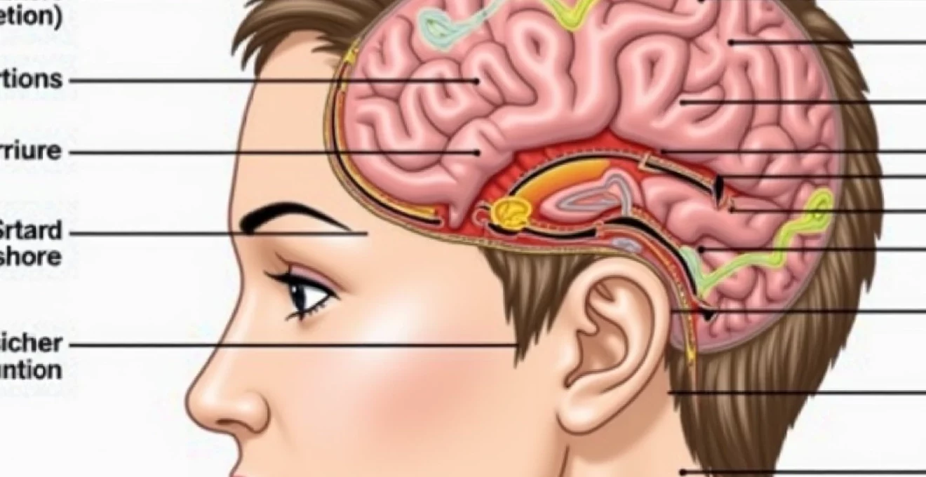
The scalp receives sensory innervation from multiple nerve sources, creating a complex network that can be disrupted at various anatomical levels. When these neural pathways are compromised, patients experience altered sensation ranging from mild tingling to complete numbness. The pathophysiology underlying scalp numbness involves either direct nerve damage, compression, or dysfunction of the sensory processing centres in the central nervous system. Peripheral neuropathy affecting the scalp nerves can result from mechanical compression, inflammatory processes, or metabolic disturbances that impair nerve conduction.
Trigeminal nerve distribution and sensory innervation patterns
The trigeminal nerve, specifically its ophthalmic and maxillary divisions, provides sensory innervation to the anterior two-thirds of the scalp. The ophthalmic division branches into the supraorbital, supratrochlear, and lacrimal nerves, whilst the maxillary division contributes through the zygomaticotemporal nerve. Dysfunction of these trigeminal branches can cause numbness in distinct anatomical patterns, helping clinicians localise the site of pathology. Trigeminal neuralgia, post-herpetic neuralgia, and mechanical compression at skull base foramina represent common causes of trigeminal-mediated scalp numbness.
Occipital nerve dysfunction and C2-C3 dermatome involvement
The posterior scalp receives innervation from the greater, lesser, and third occipital nerves, which arise from the medial branches of the C2 and C3 spinal nerves. These nerves traverse through the suboccipital triangle and pierce the tendinous insertions of the trapezius and splenius capitis muscles before innervating the posterior scalp. Occipital nerve entrapment commonly occurs at these muscular penetration points, particularly during periods of cervical muscle tension or following neck trauma. The resulting numbness typically follows the characteristic C2-C3 dermatome distribution, extending from the suboccipital region to the vertex of the skull.
Greater auricular nerve compromise and temporal region numbness
The greater auricular nerve, originating from the C2-C3 nerve roots, provides sensory innervation to the ear and surrounding temporal scalp region. This nerve is particularly vulnerable to injury during surgical procedures involving the neck or parotid gland, as it courses superficially over the sternocleidomastoid muscle. Compromise of the greater auricular nerve results in numbness affecting the earlobe, external auditory canal, and adjacent temporal scalp area. Patients often describe this numbness as particularly bothersome due to its impact on daily activities such as wearing earphones or glasses.
Supraorbital and supratrochlear nerve entrapment mechanisms
The supraorbital and supratrochlear nerves emerge from the orbit through their respective foramina or notches in the frontal bone, subsequently innervating the forehead and anterior scalp. These nerves are susceptible to entrapment at their bony exit points, particularly in individuals with anatomical variants such as narrow foramina or prominent brow ridges. Chronic muscle tension in the frontalis and corrugator supercilii muscles can also compress these nerves, leading to frontal scalp numbness. The condition is often exacerbated by repetitive facial expressions or prolonged frowning, making it common in individuals with chronic stress or anxiety disorders.
Primary aetiological factors in scalp sensory loss
Understanding the primary causes of scalp numbness requires a systematic approach that considers both local and systemic factors affecting nerve function. The aetiology can be broadly categorised into primary headache disorders, localised nerve pathology, and secondary causes related to underlying medical conditions. Each category presents with distinct clinical features and requires specific diagnostic approaches to ensure accurate identification and appropriate treatment.
Migraine-associated allodynia and cutaneous hypersensitivity
Migraine-associated scalp numbness often occurs as part of the broader phenomenon of cutaneous allodynia, where normally non-painful stimuli become painful or uncomfortable. During migraine episodes, central sensitisation of trigeminal sensory neurons leads to altered processing of sensory information, resulting in abnormal scalp sensations. Allodynia typically manifests as scalp tenderness, numbness, or hypersensitivity to light touch, making activities such as hair brushing or wearing hats extremely uncomfortable. This phenomenon affects approximately 60-80% of migraine sufferers and often serves as an indicator of migraine progression and central sensitisation.
Tension-type headache with pericranial muscle tenderness
Tension-type headaches frequently involve sustained contraction of pericranial muscles, including the frontalis, temporalis, and suboccipital muscle groups. This chronic muscle tension can lead to compression of sensory nerves as they traverse through muscular planes, resulting in localised scalp numbness. The numbness typically correlates with areas of maximal muscle tension and may fluctuate with headache intensity. Patients often report a “tight band” sensation around the head, accompanied by areas of reduced sensation that correspond to nerve compression sites.
Occipital neuralgia and arnold’s neuralgia differentiation
Occipital neuralgia represents a distinct headache disorder characterised by sharp, shooting pain in the distribution of the occipital nerves, often accompanied by scalp numbness between episodes. The condition results from irritation or injury to the greater, lesser, or third occipital nerves and can be distinguished from other headache types by its characteristic pain quality and distribution. Arnold’s neuralgia , affecting the auriculotemporal nerve, presents with similar symptoms but involves the temporal region and ear. Both conditions may cause persistent numbness in their respective nerve distributions, particularly following acute episodes of neuralgic pain.
Post-herpetic neuralgia following herpes zoster ophthalmicus
Herpes zoster ophthalmicus, affecting the ophthalmic division of the trigeminal nerve, can result in persistent scalp numbness as part of post-herpetic neuralgia. The condition typically follows the acute vesicular eruption and represents ongoing nerve dysfunction characterised by altered sensation, allodynia, and hyperalgesia. The numbness often occurs in a distinct dermatomal pattern corresponding to the original zoster distribution and may persist for months or years following the initial infection. This form of scalp numbness is particularly challenging to treat and often requires multimodal therapeutic approaches.
Systemic medical conditions causing scalp hypoesthesia
Scalp numbness can serve as an early indicator of various systemic medical conditions that affect peripheral nerve function or central nervous system processing. Diabetes mellitus represents one of the most common systemic causes, with diabetic peripheral neuropathy affecting small nerve fibres responsible for scalp sensation. The mechanism involves chronic hyperglycaemia leading to nerve damage through advanced glycation end products, oxidative stress, and microvascular dysfunction. Patients with diabetic neuropathy may experience scalp numbness alongside more typical symptoms affecting the hands and feet.
Multiple sclerosis frequently presents with sensory disturbances, including scalp numbness, as a result of demyelinating lesions affecting sensory pathways. The numbness in MS patients is often episodic and may be accompanied by other neurological symptoms such as vision changes, weakness, or coordination difficulties. Central sensitisation in MS can also lead to altered pain processing, making some patients experience scalp hypersensitivity rather than numbness. The location and pattern of scalp involvement can provide important clues about the anatomical location of demyelinating lesions.
Early recognition of scalp numbness as a potential manifestation of systemic disease can significantly improve patient outcomes through prompt diagnosis and treatment initiation.
Autoimmune conditions such as systemic lupus erythematosus, Sjögren’s syndrome, and vasculitis can cause scalp numbness through various mechanisms including small vessel disease, direct nerve infiltration, or secondary effects of chronic inflammation. These conditions often present with additional systemic symptoms that help guide diagnosis, but scalp numbness may occasionally be the presenting feature. Thyroid disorders, particularly hypothyroidism, can also contribute to peripheral neuropathy affecting scalp sensation through effects on nerve metabolism and myelination.
Iatrogenic causes and surgical complications
Surgical procedures involving the head and neck region carry inherent risks of nerve injury that can result in scalp numbness. Neurosurgical procedures, particularly those involving craniotomy or skull base surgery, may damage sensory nerves during tissue manipulation or bone work. The supraorbital and supratrochlear nerves are particularly vulnerable during frontal craniotomy approaches, whilst occipital nerve injury can occur during suboccipital procedures. Facelift surgery and other cosmetic procedures may also inadvertently damage branches of the trigeminal nerve or greater auricular nerve, leading to persistent scalp numbness in the distribution of the affected nerve.
Radiation therapy to the head and neck region can cause delayed onset scalp numbness through direct nerve injury or secondary fibrosis affecting nerve function. The numbness typically develops weeks to months following radiation completion and may be permanent depending on the radiation dose and nerve involvement. Chemotherapy agents, particularly platinum-based compounds and taxanes, can cause peripheral neuropathy that may affect scalp sensation as part of a more generalised sensory disturbance. The numbness usually develops gradually during treatment and may improve following chemotherapy completion, though some patients experience persistent symptoms.
Dental procedures, especially those involving nerve blocks or oral surgery, can occasionally result in scalp numbness through injury to branches of the trigeminal nerve. Third molar extraction and endodontic procedures carry particular risk due to the proximity of these procedures to major nerve trunks. Local anaesthetic injections can also cause temporary or permanent nerve damage, particularly when performed in anatomically variant locations or with excessive injection pressure.
Comprehensive clinical assessment protocols
Thorough clinical evaluation of scalp numbness requires a systematic approach that combines detailed history taking with comprehensive neurological examination. The assessment should begin with characterisation of the numbness, including its onset, distribution, associated symptoms, and potential triggering factors. Clinicians should enquire about recent trauma, surgical procedures, infections, or new medications that might contribute to nerve dysfunction. The quality of the numbness—whether it is complete loss of sensation, reduced sensation, or altered sensation—provides important diagnostic clues about the underlying pathophysiology.
Monofilament testing and quantitative sensory testing applications
Monofilament testing provides an objective method for assessing light touch sensation across different areas of the scalp. Using graded monofilaments, clinicians can quantify sensory thresholds and map areas of reduced sensation with precision. Quantitative sensory testing (QST) offers more sophisticated assessment of multiple sensory modalities including vibration, thermal sensation, and pain thresholds. These tests are particularly valuable for documenting baseline function and monitoring response to treatment over time. QST protocols specifically designed for trigeminal nerve assessment can help localise dysfunction to specific nerve branches.
Nerve conduction studies and electromyography interpretation
Electrodiagnostic testing plays a crucial role in evaluating suspected peripheral nerve involvement in scalp numbness. Nerve conduction studies can assess the function of accessible nerves such as the supraorbital branch of the trigeminal nerve, though technical limitations exist for testing all scalp nerves. Electromyography of relevant muscles innervated by mixed nerves can provide additional information about nerve function. Blink reflex testing offers a specific method for evaluating trigeminal nerve function and can detect subclinical abnormalities that might not be apparent on clinical examination alone.
MRI neurography and High-Resolution ultrasound imaging
Advanced imaging techniques provide valuable anatomical information about nerve pathways and potential sites of compression or injury. MRI neurography uses specialised sequences to visualise peripheral nerves and can identify nerve swelling, compression, or discontinuity. High-resolution ultrasound has emerged as a valuable tool for assessing superficial nerves such as the occipital and supraorbital nerves, allowing real-time visualisation during dynamic manoeuvres. These imaging modalities are particularly useful for identifying treatable causes of nerve compression and planning targeted interventions.
Differential diagnosis using weber and rinne testing
Although primarily used for hearing assessment, Weber and Rinne testing can provide valuable information when scalp numbness involves the auricular region or is associated with auditory symptoms. Abnormal results may suggest involvement of the facial nerve or acoustic neuroma, which can cause secondary effects on surrounding sensory nerves. Corneal reflex testing specifically evaluates trigeminal nerve function and should be performed when frontal scalp numbness is present, as abnormalities may indicate more extensive trigeminal involvement requiring urgent investigation.
Evidence-based treatment modalities and prognosis
Treatment of scalp numbness must be tailored to the underlying aetiology and may involve pharmacological, interventional, or surgical approaches. For neuropathic causes, anticonvulsants such as gabapentin or pregabalin serve as first-line treatments, with typical starting doses of 300mg daily for gabapentin or 75mg twice daily for pregabalin. Tricyclic antidepressants like amitriptyline or nortriptyline provide alternative options, particularly when sleep disturbance accompanies the numbness. Topical treatments including lidocaine patches or capsaicin cream may offer localised relief without systemic side effects.
Interventional approaches include nerve blocks using local anaesthetics or corticosteroids, which can provide both diagnostic and therapeutic benefit. Greater occipital nerve blocks are particularly effective for posterior scalp numbness, whilst supraorbital nerve blocks address frontal scalp symptoms. Botulinum toxin injections have shown promise for treating muscle tension-related scalp numbness and can provide relief lasting 3-6 months. For refractory cases, neurolytic procedures or surgical nerve decompression may be considered, though these carry additional risks and require careful patient selection.
The prognosis for scalp numbness varies significantly depending on the underlying cause, with acute inflammatory conditions typically showing better recovery potential than chronic degenerative processes.
Non-pharmacological treatments play an important complementary role in managing scalp numbness. Physical therapy focusing on cervical spine mobility and posture correction can address musculoskeletal contributors to nerve compression. Stress reduction techniques and cognitive behavioural therapy may be particularly beneficial for patients with tension-type headache-related scalp numbness. Transcutaneous electrical nerve stimulation (TENS) provides a non-invasive option that some patients find helpful for managing chronic symptoms. Regular follow-up is essential to monitor treatment response and adjust therapy as needed, with most patients experiencing some degree of improvement within 4-6 weeks of initiating appropriate treatment.