Purple visual phenomena represent one of the most intriguing aspects of ocular and neurological health, affecting millions of individuals worldwide with varying degrees of intensity and clinical significance. These chromatic disturbances can manifest as fleeting purple spots, persistent coloured overlays, or complex visual aberrations that significantly impact daily functioning. Understanding the underlying mechanisms behind purple vision requires a comprehensive examination of both physiological and pathological processes affecting the visual system. From benign entoptic phenomena to serious retinal pathologies, purple visual symptoms demand careful evaluation to distinguish between harmless perceptual quirks and potentially sight-threatening conditions.
The complexity of purple visual manifestations lies in their diverse aetiological origins, ranging from simple mechanical pressure on the eyeball to sophisticated neurological disorders affecting visual cortex processing. Modern ophthalmology and neurology have made significant strides in understanding these phenomena, yet many patients continue to experience anxiety when confronted with unexpected purple visual disturbances. The key to appropriate management lies in recognising the distinguishing characteristics of different purple visual symptoms and understanding when immediate medical intervention becomes necessary.
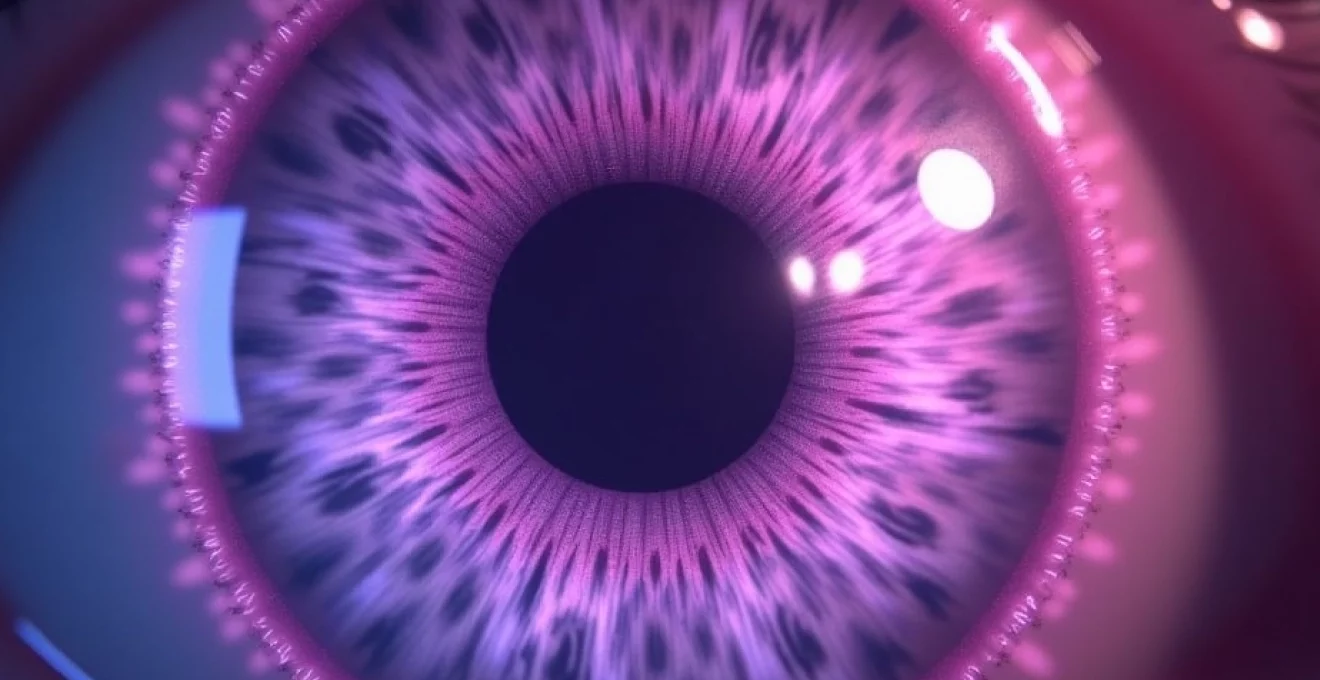
Understanding purple visual phenomena: chromatic aberrations and photopsia
Purple visual phenomena encompass a broad spectrum of chromatic disturbances that can occur within the visual system, each with distinct characteristics and underlying mechanisms. These manifestations often represent the brain’s interpretation of abnormal electrical activity within the visual pathway, from the retinal photoreceptors to the occipital cortex. The perception of purple specifically occurs when the visual system processes wavelengths around 380-450 nanometres, though pathological conditions can create artificial purple sensations even in the absence of actual purple light stimuli.
The physiological basis for purple vision involves complex interactions between different types of photoreceptors and their corresponding neural pathways. When these systems become disrupted through mechanical pressure, chemical imbalance, or pathological processes, the resulting electrical signals can be misinterpreted by the brain as purple light. This misinterpretation explains why patients often report seeing purple spots or flashes during migraine auras, following head trauma, or as a result of certain medications affecting neurotransmitter balance.
Entoptic phenomena and retinal light sensitivity
Entoptic phenomena represent normal visual experiences that become noticeable under specific circumstances, often involving the perception of structures within the eye itself. These phenomena can manifest as purple-tinted visual disturbances, particularly when viewing bright backgrounds such as clear blue skies or uniformly illuminated surfaces. The entoptic phenomenon most commonly associated with purple visual experiences involves the visualisation of white blood cells moving through retinal capillaries, which can appear as small, rapidly moving purple or blue-white spots in the central visual field.
The mechanism behind entoptic purple vision relates to the differential absorption and scattering of light by various intraocular structures. When light passes through the vitreous gel, lens, and other transparent media within the eye, certain wavelengths become preferentially scattered or absorbed, potentially creating the appearance of purple-tinted moving objects. This phenomenon becomes more pronounced with age as the vitreous undergoes natural liquefaction processes, leading to increased light scattering and more prominent entoptic experiences.
Phosphene-induced purple flashes during eye pressure
Phosphenes represent one of the most common causes of purple visual phenomena, occurring when mechanical pressure is applied to the eyeball or when electrical activity stimulates the visual system in the absence of light. These pressure-induced purple flashes typically manifest as brief, brilliant spots or geometric patterns that can range from simple dots to complex kaleidoscopic formations. The purple coloration often results from the simultaneous activation of multiple photoreceptor types, creating a mixed signal that the brain interprets as purple light.
Mechanical phosphenes occur most frequently when individuals rub their eyes, experience sudden changes in intraocular pressure, or sustain minor trauma to the eye. The resulting purple flashes usually last only seconds but can be quite vivid and alarming to those unfamiliar with the phenomenon. Understanding that these experiences represent normal physiological responses to pressure can provide reassurance, though persistent or spontaneous phosphenes may indicate underlying pathology requiring medical evaluation.
Blue field entoptic phenomenon and leucocyte visualization
The blue field entoptic phenomenon represents a fascinating aspect of normal vision that can sometimes be perceived as purple-tinted moving spots, particularly when observing bright blue backgrounds. This phenomenon involves the direct visualisation of white blood cells moving through retinal capillaries, creating small, rapidly moving bright spots that dart across the visual field. While these spots typically appear white or pale blue, certain lighting conditions and individual variations in colour perception can cause them to appear purple or violet.
This phenomenon becomes most apparent when looking at uniform blue backgrounds, such as clear skies, and can be enhanced by focusing attention on the peripheral visual field. The moving spots typically follow curved paths corresponding to the retinal vascular architecture and move at speeds consistent with normal blood flow. Recognition of this normal phenomenon prevents unnecessary anxiety when individuals first notice these purple-tinted moving objects in their vision, though sudden increases in the number or intensity of these spots should prompt medical evaluation.
Chromatopsia: Purple-Tinted vision disturbances
Chromatopsia refers to the pathological perception of colour tints overlaying normal vision, with purple chromatopsia representing a specific subtype that can significantly impact visual function and quality of life. This condition can result from various causes, including medication side effects, retinal pathology, or neurological disorders affecting colour processing centres in the brain. Purple chromatopsia often presents as a persistent purple overlay across the entire visual field, though it can also manifest as intermittent episodes of purple-tinted vision.
The differential diagnosis of purple chromatopsia requires careful consideration of medication history, particularly drugs known to affect colour perception such as certain antimalarials, cardiac glycosides, and psychoactive substances. Retinal causes of purple chromatopsia include cone dystrophies, advanced macular degeneration, and certain inflammatory conditions affecting the macula. Neurological causes may involve lesions affecting the visual cortex or pathways involved in colour processing, highlighting the importance of comprehensive evaluation when patients report persistent purple visual tinting.
Ocular pathologies causing purple vision manifestations
Numerous ocular pathologies can manifest with purple visual symptoms, ranging from benign age-related changes to serious sight-threatening conditions requiring immediate intervention. These pathological processes typically involve disruption of normal retinal architecture, vitreous transparency, or intraocular pressure regulation, leading to abnormal light transmission and processing within the eye. The appearance of purple visual phenomena in the context of ocular disease often represents secondary effects of primary pathological processes rather than direct purple light generation.
Understanding the relationship between specific ocular pathologies and purple visual manifestations requires knowledge of normal visual physiology and how disease processes disrupt these mechanisms. For instance, retinal haemorrhages can create areas of altered light transmission that appear purple under certain lighting conditions, while vitreous opacities can scatter light in ways that produce purple-tinted visual disturbances. The timing, location, and associated symptoms of purple visual phenomena provide crucial diagnostic clues for identifying underlying ocular pathology.
Retinal detachment and posterior vitreous detachment symptoms
Retinal detachment and posterior vitreous detachment represent serious ocular emergencies that can manifest with purple visual symptoms among their constellation of presenting signs. During the initial stages of vitreous detachment, mechanical traction on the retina can stimulate photoreceptors inappropriately, leading to the perception of flashing lights that may appear purple, white, or multicoloured. These photopsic phenomena often occur in the peripheral visual field and may be accompanied by the sudden onset of new floaters or curtain-like visual field defects.
The progression from posterior vitreous detachment to retinal detachment can be accompanied by increasingly prominent purple or coloured visual disturbances as retinal architecture becomes progressively disrupted. Early recognition of these symptoms is crucial for preventing permanent visual loss, as prompt surgical intervention can often restore retinal attachment and preserve vision. Patients experiencing new onset purple flashes, particularly when accompanied by floaters or visual field defects, require urgent ophthalmological evaluation to rule out retinal detachment.
Migraine aura scintillations and visual cortex disturbances
Migraine aura represents one of the most common causes of purple visual phenomena, affecting approximately 25-30% of migraine sufferers with characteristic visual disturbances that can be both dramatic and frightening. The classic migraine aura typically begins as a small, scintillating purple or multicoloured spot near the centre of vision, which gradually expands into a crescent-shaped area of flashing lights bordered by a blind spot. This phenomenon, known as a scintillating scotoma, results from cortical spreading depression affecting the primary visual cortex.
The purple coloration commonly associated with migraine auras reflects the brain’s interpretation of abnormal electrical activity spreading across the visual cortex in a characteristic wave-like pattern. These visual disturbances typically last 15-30 minutes and may be followed by the onset of headache, though some individuals experience visual aura without subsequent pain.
The relationship between cortical spreading depression and purple visual phenomena demonstrates the complex interplay between neurological and visual processes in migraine pathophysiology.
Diabetic retinopathy advanced stages and haemorrhaging
Advanced diabetic retinopathy can present with purple visual manifestations related to retinal haemorrhaging, neovascularisation, and associated inflammatory processes. As diabetes progresses and affects retinal blood vessels, areas of bleeding can create purple or reddish-purple spots within the visual field, particularly noticeable against bright backgrounds. These haemorrhages can vary in size from small dot-like lesions to larger blot haemorrhages that significantly impact visual acuity and contrast sensitivity.
The progression of diabetic retinopathy from background changes to proliferative disease can be accompanied by increasingly prominent purple visual disturbances as neovascularisation and associated bleeding compromise normal retinal architecture. Regular monitoring and early intervention remain crucial for preventing vision-threatening complications, with purple visual symptoms often serving as early warning signs of disease progression requiring intensified medical management or surgical intervention.
Macular degeneration metamorphopsia effects
Age-related macular degeneration can manifest with purple visual disturbances, particularly during the intermediate and advanced stages when retinal pigment epithelium dysfunction and choroidal neovascularisation significantly impact colour perception and visual processing. The accumulation of drusen deposits and associated inflammatory changes can alter light transmission through the macula, potentially creating areas that appear purple-tinted or cause surrounding colours to appear shifted towards the purple spectrum.
Metamorphopsia, the distortion of straight lines and geometric shapes characteristic of macular degeneration, can be accompanied by colour disturbances that manifest as purple-tinted areas within the central visual field. These changes often develop gradually and may initially be subtle, making regular monitoring with tools such as Amsler grids essential for early detection. The relationship between macular degeneration progression and purple visual symptoms underscores the importance of comprehensive retinal evaluation when patients report new-onset colour disturbances.
Cataracts-related chromatic dispersion and light scattering
Cataract formation can contribute to purple visual phenomena through altered light transmission and increased intraocular light scattering that affects colour perception and visual quality. As lens proteins undergo age-related changes and become increasingly opacified, certain wavelengths of light may be preferentially scattered or absorbed, potentially creating the appearance of purple-tinted vision or purple halos around light sources. These chromatic effects often become more pronounced in low-light conditions or when viewing bright lights against dark backgrounds.
The relationship between cataract density, location, and purple visual symptoms varies significantly among individuals, with posterior subcapsular cataracts often producing more dramatic visual disturbances than nuclear or cortical cataracts. Understanding the role of cataracts in purple visual phenomena is important for appropriate surgical planning and patient counselling, as cataract extraction typically resolves chromatic aberrations and restores normal colour perception.
Neurological conditions triggering purple visual disturbances
Neurological conditions affecting the visual pathway from the optic nerves to the occipital cortex can produce complex purple visual disturbances that often differ significantly from those caused by primary ocular pathology. These neurological purple visual phenomena typically result from abnormal electrical activity, structural lesions, or biochemical imbalances affecting neurons involved in visual processing. The characteristics of neurologically-mediated purple vision often provide important diagnostic clues about the location and nature of underlying pathology.
The complexity of neurological purple visual disturbances reflects the sophisticated nature of central visual processing, where multiple brain regions collaborate to create coherent visual experiences. When these systems become disrupted by disease, injury, or biochemical imbalance, the resulting visual phenomena can be both varied and unpredictable. Understanding the neurological basis of purple visual disturbances requires knowledge of visual pathway anatomy and the specific ways different neurological conditions can affect visual processing centres.
Occipital lobe seizures and temporal lobe epilepsy
Seizure activity affecting the occipital lobe can produce dramatic purple visual phenomena as abnormal electrical discharges spread through visual processing centres in the brain. These seizure-related visual disturbances, known as visual seizures or occipital seizures, can manifest as brilliant purple lights, geometric patterns, or complex visual hallucinations that may be accompanied by other neurological symptoms. The purple coloration often reflects the simultaneous activation of multiple visual processing pathways during seizure activity.
Temporal lobe epilepsy can also produce purple visual disturbances, particularly when seizure activity spreads to involve visual association areas or when complex partial seizures affect consciousness and visual perception. These episodes may be accompanied by other temporal lobe seizure symptoms such as déjà vu experiences, emotional changes, or automatisms. Recognition of seizure-related purple visual phenomena is crucial for appropriate neurological evaluation and treatment, as these symptoms may represent the only manifestation of underlying epileptic activity.
Cortical spreading depression in migraine pathophysiology
Cortical spreading depression represents the primary mechanism underlying migraine aura, including the characteristic purple visual phenomena experienced by many migraine sufferers. This wave of neuronal depolarisation spreads across the visual cortex at a rate of approximately 2-3 millimetres per minute, creating the gradually expanding purple or multicoloured visual disturbances typical of migraine aura. The phenomenon demonstrates the intricate relationship between neuronal electrical activity and visual perception.
The progression of cortical spreading depression through different areas of the visual cortex explains the characteristic evolution of migraine aura symptoms, from initial purple spots to expanding crescents of scintillating lights followed by areas of visual field depression.
Understanding cortical spreading depression provides insights into both the mechanisms of migraine aura and potential therapeutic targets for preventing these debilitating visual disturbances.
Recent research has revealed that cortical spreading depression may also occur in other neurological conditions, expanding our understanding of purple visual phenomena beyond traditional migraine contexts.
Transient ischaemic attacks affecting visual processing
Transient ischaemic attacks affecting the posterior circulation can produce temporary purple visual disturbances as reduced blood flow compromises visual processing centres in the occipital cortex and brainstem. These ischaemic episodes may manifest as purple-tinted vision, purple spots, or more complex visual disturbances that typically resolve within minutes to hours as normal blood flow is restored. The temporary nature of these symptoms reflects the reversible dysfunction of neural tissue during periods of reduced oxygen and glucose supply.
The pattern and progression of purple visual symptoms during transient ischaemic attacks can provide important diagnostic information about the location and severity of vascular compromise. Bilateral purple visual disturbances often suggest involvement of the posterior cerebral artery circulation, while unilateral symptoms may indicate more localised vascular pathology. Early recognition and appropriate evaluation of these symptoms are crucial for stroke prevention and optimisation of vascular risk factors.
Multiple sclerosis optic neuritis manifestations
Multiple sclerosis can produce purple visual disturbances through various mechanisms, including optic neuritis, demyelinating lesions affecting visual pathways, and Uhthoff’s phenomenon related to temperature-sensitive conduction block. Optic neuritis, one of the most common presenting features of multiple sclerosis, can cause colour desaturation and abnormal colour perception that may manifest as purple-tinted vision or difficulty distinguishing purple from other colours. These symptoms often accompany central vision loss, eye pain, and contrast sensitivity reduction characteristic of optic neuritis.
Demyelinating lesions affecting the visual cortex or optic radiations can produce more complex purple visual phenomena, including visual field defects with purple-tinted borders or persistent purple overlays affecting portions of the visual field. The relationship between multiple
sclerosis manifestations and purple visual disturbances highlights the importance of comprehensive neurological evaluation when these symptoms occur in the context of other demyelinating disease features such as fatigue, cognitive changes, or motor dysfunction.
Pharmacological and toxic causes of purple vision
Numerous medications and toxic substances can induce purple visual disturbances through various mechanisms affecting retinal function, neurotransmitter balance, or visual processing centres in the brain. These pharmacologically-induced visual phenomena often represent dose-dependent side effects that may resolve with medication adjustment or discontinuation, though some effects can persist even after cessation of the offending agent. Understanding the relationship between specific medications and purple visual symptoms is crucial for appropriate clinical management and patient counselling.
Antimalarial medications, particularly chloroquine and hydroxychloroquine, represent some of the most well-documented causes of purple visual disturbances through their effects on retinal pigment epithelium function and photoreceptor cell integrity. These medications can accumulate in retinal tissues over time, leading to progressive visual field defects and colour vision abnormalities that may include purple chromatopsia or difficulty distinguishing purple from other colours. The irreversible nature of advanced antimalarial retinopathy underscores the importance of regular ophthalmological monitoring during treatment with these agents.
Cardiac glycosides such as digoxin can produce characteristic purple or yellow visual tinting as a result of their effects on cellular sodium-potassium pumps within retinal tissues. These chromatic disturbances often serve as early indicators of digoxin toxicity and may be accompanied by other symptoms such as nausea, fatigue, or cardiac rhythm disturbances. Psychoactive substances, including certain antidepressants, antipsychotics, and recreational drugs, can also induce purple visual phenomena through their effects on neurotransmitter systems involved in visual processing and colour perception.
Diagnostic procedures for purple visual symptoms
Comprehensive evaluation of purple visual symptoms requires a systematic approach incorporating detailed history taking, thorough physical examination, and appropriate diagnostic testing to differentiate between benign phenomena and serious underlying pathology. The diagnostic process must consider the temporal characteristics of symptoms, associated features, medication history, and relevant medical conditions that might predispose to visual disturbances. Modern ophthalmological and neurological diagnostic techniques provide sophisticated tools for investigating the underlying causes of purple visual phenomena.
The initial evaluation should focus on characterising the specific nature of purple visual symptoms, including their location within the visual field, duration, triggers, and associated symptoms such as headache, eye pain, or neurological deficits. This information helps guide subsequent diagnostic testing and determines the urgency of further evaluation. Understanding whether symptoms are monocular or binocular, central or peripheral, and constant or intermittent provides crucial diagnostic clues about the likely anatomical location of pathology.
Fundoscopy and optical coherence tomography examination
Direct and indirect fundoscopy remain fundamental components of evaluating purple visual symptoms, allowing detailed examination of retinal architecture, vascular patterns, and potential pathological changes that might explain visual disturbances. The fundoscopic examination can identify retinal haemorrhages, exudates, cotton wool spots, or other abnormalities that might appear purple-tinted or contribute to chromatic visual disturbances. Careful attention to the macula, optic nerve, and peripheral retina helps identify specific pathological processes that require targeted treatment.
Optical coherence tomography provides high-resolution cross-sectional imaging of retinal layers, enabling detection of subtle structural abnormalities that might not be visible during clinical examination. This technology proves particularly valuable for identifying macular oedema, retinal pigment epithelium changes, or vitreoretinal interface abnormalities that could contribute to purple visual phenomena. Advanced OCT imaging techniques, including angiography and en face imaging, offer additional insights into retinal vascular perfusion and structural integrity that may be relevant to purple visual symptoms.
Visual field testing using humphrey perimetry
Automated perimetry using instruments such as the Humphrey visual field analyser provides objective assessment of visual field integrity and can identify scotomas or visual field defects associated with purple visual phenomena. These tests prove particularly valuable when purple visual symptoms are described as occurring in specific areas of the visual field or when neurological causes are suspected. The pattern and characteristics of visual field defects can help localise pathology within the visual pathway from retina to occipital cortex.
Specialised visual field testing protocols, including blue-on-yellow perimetry and frequency-doubling technology, may be employed to detect early changes in specific retinal cell populations that might contribute to colour vision disturbances. These advanced techniques can identify functional abnormalities before structural changes become apparent, enabling earlier intervention and better visual outcomes. The correlation between visual field defects and subjective purple visual symptoms helps validate patient complaints and guides treatment decisions.
Electroretinography for retinal function assessment
Electroretinography provides objective measurement of retinal electrical activity and can identify functional abnormalities in specific retinal cell populations that might contribute to purple visual disturbances. This testing proves particularly valuable when investigating suspected retinal dystrophies, toxic retinopathies, or inflammatory conditions affecting retinal function. The pattern of electroretinographic abnormalities can help distinguish between different causes of visual dysfunction and guide appropriate treatment strategies.
Multifocal electroretinography enables localised assessment of retinal function across different areas of the posterior pole, providing detailed information about macular function that may be relevant to central purple visual symptoms. Pattern electroretinography specifically evaluates retinal ganglion cell function and may be useful when investigating optic nerve dysfunction or central nervous system causes of visual disturbances. These sophisticated electrophysiological techniques complement clinical examination and imaging studies in the comprehensive evaluation of purple visual phenomena.
MRI neuroimaging for cortical lesion detection
Magnetic resonance imaging of the brain and orbits provides detailed visualisation of the visual pathway from optic nerves to occipital cortex, enabling identification of structural abnormalities that might cause purple visual disturbances. High-resolution MRI sequences can detect demyelinating lesions, mass lesions, vascular abnormalities, or inflammatory changes affecting visual processing centres in the brain. The addition of contrast enhancement may reveal active inflammatory processes or vascular abnormalities requiring specific treatment.
Functional MRI techniques can provide insights into visual cortex activity and may help identify abnormal activation patterns associated with purple visual phenomena. These advanced imaging modalities prove particularly valuable when investigating complex visual disturbances that cannot be explained by ophthalmological examination alone. The correlation between MRI findings and clinical symptoms guides therapeutic decisions and helps predict visual outcomes.
Treatment protocols and management strategies
Management of purple visual symptoms requires a targeted approach based on accurate diagnosis of the underlying cause, with treatment strategies ranging from simple reassurance for benign phenomena to urgent surgical intervention for sight-threatening conditions. The therapeutic approach must consider not only the specific pathological process involved but also patient factors such as age, overall health status, visual function requirements, and treatment preferences. Successful management often requires coordination between ophthalmologists, neurologists, and other specialists to address complex multisystem conditions.
Treatment protocols for purple visual symptoms can be broadly categorised into medical management, surgical intervention, and supportive care measures aimed at optimising visual function and quality of life. Medical management may involve systemic medications, topical treatments, or lifestyle modifications designed to address underlying pathological processes or reduce symptom severity. Surgical interventions range from outpatient laser procedures to complex intraocular surgeries depending on the specific condition identified.
For migraine-related purple visual phenomena, preventive treatments may include beta-blockers, calcium channel blockers, anticonvulsants, or newer targeted therapies such as CGRP antagonists that can significantly reduce both the frequency and severity of visual aura episodes. Acute treatment strategies focus on early intervention with appropriate analgesics or triptans, though these medications should be used judiciously to avoid medication overuse complications. Lifestyle modifications including trigger identification and avoidance, stress management, and sleep hygiene optimisation play crucial roles in comprehensive migraine management.
When purple visual symptoms result from retinal pathology such as diabetic retinopathy or macular degeneration, treatment protocols may involve anti-VEGF injections, corticosteroids, laser photocoagulation, or surgical procedures designed to preserve or restore retinal function. These treatments require careful monitoring and often involve repeated interventions over extended periods to maintain visual stability. Patient education about disease progression, treatment expectations, and potential complications ensures informed decision-making and optimal treatment adherence.
Neurological causes of purple visual disturbances may require anticonvulsant medications for seizure-related symptoms, immunomodulatory therapies for inflammatory conditions such as multiple sclerosis, or vascular risk factor modification for cerebrovascular causes. The complexity of neurological treatments necessitates close collaboration with neurology specialists and regular monitoring for treatment efficacy and potential side effects. Rehabilitation strategies including visual therapy, adaptive techniques, and assistive technologies help patients maintain independence and quality of life despite persistent visual symptoms.
For medication-induced purple visual phenomena, management typically involves dose reduction, medication substitution, or gradual discontinuation under medical supervision when clinically appropriate. However, some conditions requiring long-term treatment with potentially retinotoxic medications necessitate careful risk-benefit analysis and regular monitoring to detect early signs of visual toxicity. Patient counselling about the importance of regular ophthalmological examinations and prompt reporting of new visual symptoms ensures early detection and intervention for medication-related complications.