The presence of gas bubbles in the eye following vitreoretinal surgery creates a fascinating optical phenomenon that fundamentally alters how patients perceive their visual world. These therapeutic gas injections, whilst essential for successful retinal reattachment and macular hole repair, introduce complex light reflection patterns that can be both bewildering and concerning for patients. Understanding the optical physics behind these reflections helps demystify the visual experiences patients encounter during their recovery period. The interplay between gas bubble properties and intraocular light transmission creates distinctive visual effects that serve as important indicators of healing progress whilst simultaneously presenting unique challenges for patient adaptation.
Understanding gas bubble formation during vitreoretinal surgery procedures
Vitreoretinal surgery frequently employs gas tamponade techniques to achieve optimal surgical outcomes, particularly in cases involving retinal detachment, macular holes, and complex vitreous pathology. The introduction of gas bubbles into the vitreous cavity serves multiple therapeutic purposes, acting as an internal splint to maintain retinal positioning whilst tissues heal. The choice of gas agent significantly influences both the duration of tamponade and the optical characteristics that patients experience during recovery. Modern surgical practice predominantly utilises three main tamponade agents: sulfur hexafluoride, perfluoropropane, and silicone oil, each presenting distinct optical properties that affect light transmission and reflection patterns.
The formation of these therapeutic gas bubbles occurs through precise injection techniques that replace the natural vitreous gel with carefully measured volumes of expansile gases. This replacement fundamentally alters the refractive index within the eye , creating sharp optical interfaces where light undergoes dramatic changes in transmission behaviour. The gas-aqueous interface becomes a critical optical boundary that generates the characteristic reflections patients observe. Understanding these formation mechanisms helps predict the visual phenomena patients will encounter and provides insight into the timeline for visual recovery.
Sulfur hexafluoride (SF6) gas properties and refractive index characteristics
Sulfur hexafluoride represents the most commonly utilised short-acting gas in vitreoretinal surgery, with expansion properties that reach maximum volume within 24-48 hours post-injection. The refractive index of SF6 gas approximates 1.0005, creating a substantial optical interface when positioned against aqueous humour with its refractive index of 1.336. This significant difference in refractive indices generates pronounced reflection effects that patients describe as “spirit level” phenomena or “underwater” vision quality. The expansion characteristics of SF6 mean that initial injection volumes of 0.3-0.5ml can expand to fill 80-90% of the vitreous cavity , maximising the surface area available for light reflection.
Perfluoropropane (C3F8) gas bubble dynamics and optical behaviour
Perfluoropropane offers extended tamponade duration, typically persisting for 6-8 weeks compared to SF6’s 2-3 week lifespan. The longer molecular chains in C3F8 result in slower absorption rates and more prolonged optical effects for patients. C3F8 bubbles maintain their reflective properties throughout their extended presence , creating consistent visual disturbances that require greater patient adaptation strategies. The optical behaviour of C3F8 bubbles demonstrates more pronounced magnification effects when patients attempt to view objects through the gas medium, with magnification factors reaching 1.3-1.5 times normal size depending on viewing distance and bubble position.
Silicone oil interface effects on light transmission and reflection
Silicone oil tamponade presents unique optical challenges due to its persistent presence and distinct refractive properties. With a refractive index of approximately 1.404, silicone oil creates different reflection patterns compared to gas bubbles whilst maintaining greater optical clarity for patients. The oil-aqueous interface generates less dramatic reflection effects but introduces subtle optical aberrations that affect colour perception and contrast sensitivity. Patients with silicone oil often report improved functional vision compared to gas-filled eyes , though they may notice slight colour desaturation and reduced depth perception until oil removal.
Pneumatic retinopexy gas injection techniques and visual consequences
Pneumatic retinopexy involves office-based gas injection procedures that create immediate and dramatic visual changes for patients. The injection technique typically introduces 0.3-0.5ml of expansile gas into the mid-vitreous cavity, where it rapidly expands to create therapeutic tamponade pressure against detached retinal tissue. The immediate visual consequences include complete visual obstruction in the injected area , with patients reporting “black bubble” effects that move with eye movement. The positioning requirements following pneumatic retinopexy mean patients must maintain specific head positions to ensure the gas bubble contacts the appropriate retinal area, creating unique visual challenges as the bubble shifts with positional changes.
The key to successful pneumatic retinopexy lies in precise bubble positioning, where the gas must contact the retinal break while maintaining adequate surface tension to prevent re-detachment.
Pathophysiology of gas bubble reflections in vitreous cavity
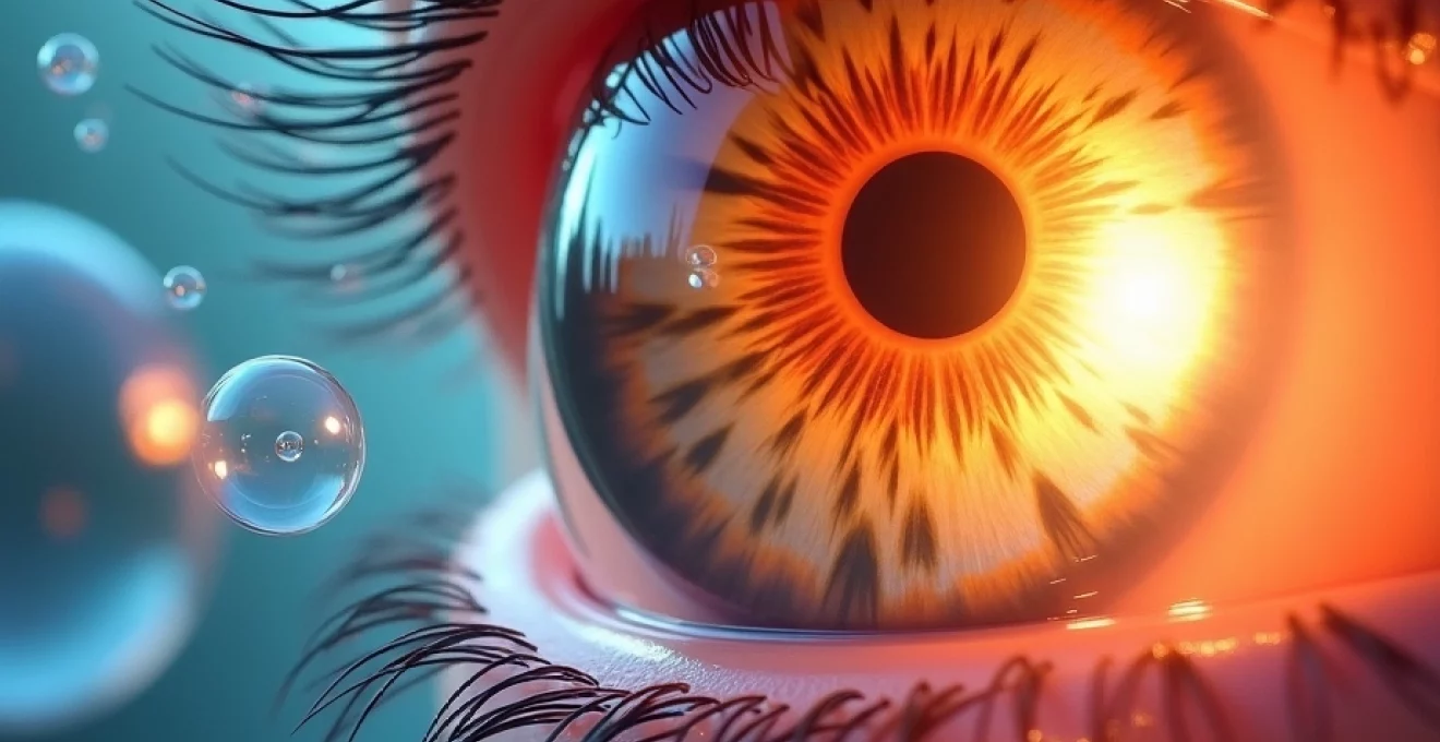
The pathophysiology underlying gas bubble reflections involves complex optical interactions between light waves, gas molecules, and surrounding ocular tissues. When light encounters the gas-aqueous interface within the vitreous cavity, multiple optical phenomena occur simultaneously, including refraction, reflection, and total internal reflection. The curvature of the gas bubble interface acts as a curved mirror system , creating both concave and convex optical surfaces depending on the viewing angle and bubble position. These curved interfaces generate varying degrees of magnification and minification effects that contribute to the disorienting visual experiences patients describe.
The physiological response to these optical disturbances involves complex neural adaptation mechanisms within the visual cortex. Patients typically experience a period of visual confusion as their brain attempts to process conflicting visual information from the treated and untreated eyes. Binocular vision becomes significantly compromised during the gas bubble phase , leading to depth perception difficulties and spatial disorientation. Understanding these pathophysiological responses helps predict patient adaptation timelines and guides appropriate counselling regarding visual expectations during recovery.
Total internal reflection mechanisms at Gas-Aqueous interface
Total internal reflection occurs when light travelling through the denser aqueous medium encounters the gas interface at angles exceeding the critical angle. For the aqueous-gas boundary, this critical angle approximates 48.6 degrees, meaning that light rays approaching at steeper angles undergo complete reflection rather than transmission. This phenomenon creates the characteristic “mirror-like” effects patients observe when viewing objects through peripheral vision whilst the gas bubble is present. The total internal reflection mechanism explains why patients often report seeing inverted or displaced images when attempting to look through or around the gas bubble.
Snell’s law applications in intraocular gas bubble optics
Snell’s law governs the refraction behaviour at the gas-aqueous interface, determining how light rays bend when transitioning between media of different refractive indices. The formula n₁sin(θ₁) = n₂sin(θ₂) describes the relationship between incident and refracted angles, where n₁ and n₂ represent the refractive indices of aqueous humour and gas respectively. The significant difference in refractive indices creates substantial ray bending , contributing to the distorted vision patients experience when looking through the gas bubble. This optical principle explains why objects viewed through the gas medium appear magnified and distorted compared to those seen through the normal aqueous-filled portions of the eye.
Fresnel reflection coefficients in Vitreous-Gas boundary interactions
Fresnel reflection coefficients determine the proportion of incident light that undergoes reflection versus transmission at the gas-aqueous interface. These coefficients vary with the angle of incidence and polarisation state of the incoming light, creating varying reflection intensities across different viewing angles. The high reflectance at certain angles contributes to the glare and light sensitivity patients frequently report during the gas bubble phase. Understanding Fresnel reflection behaviour helps explain why patients may experience varying degrees of visual disturbance depending on lighting conditions and viewing angles.
Macular pucker and epiretinal membrane impact on light scattering
Pre-existing retinal pathology, particularly macular pucker and epiretinal membranes, can significantly influence light scattering patterns in gas-filled eyes. These tissue irregularities create additional optical interfaces that interact with gas bubble reflections to produce complex visual phenomena. Patients with epiretinal membranes may experience enhanced reflection effects due to the multiple optical boundaries present within their visual axis. The combination of gas bubble reflections and retinal surface irregularities often results in increased photophobia and contrast sensitivity reduction during the recovery period.
Clinical manifestations of intraocular gas bubble visual disturbances
Patients with intraocular gas bubbles experience a spectrum of visual disturbances that evolve predictably as the bubble undergoes absorption and repositioning. The most commonly reported phenomenon involves the “spirit level” effect, where patients observe a distinct horizontal line separating clear vision above from obscured or distorted vision below. This demarcation line corresponds to the gas-aqueous interface and moves dynamically with head position changes. The clarity above this line gradually improves as inflammation subsides , whilst the area below remains optically compromised until gas absorption progresses sufficiently.
Magnification effects represent another significant clinical manifestation, with patients reporting that objects viewed through the gas bubble appear significantly enlarged compared to normal vision. This magnification can range from 1.2 to 1.8 times normal size depending on the viewing distance and bubble position within the eye. Reading becomes challenging due to the dual magnification effects from the gas bubble combined with any residual refractive changes from the surgical procedure. Patients often describe difficulty judging distances and report bumping into objects due to the altered spatial perception created by the gas bubble’s optical properties.
Reflection artifacts present as one of the most visually disturbing manifestations of gas bubble presence. Patients frequently report seeing multiple images, inverted reflections, or “ghost” images that move independently of their intended gaze direction. These artifacts result from light reflecting off the curved gas-aqueous interface before reaching the retina, creating false visual signals that the brain must attempt to interpret. The intensity and frequency of these reflection artifacts typically correlate with the size and position of the gas bubble within the vitreous cavity.
The visual experience of living with an intraocular gas bubble has been described as “looking through a funhouse mirror whilst underwater,” highlighting the complex optical distortions patients must navigate during recovery.
Light sensitivity and glare intolerance frequently accompany gas bubble presence, with patients reporting increased difficulty in bright lighting conditions. The irregular optical surfaces created by the gas bubble scatter incoming light in unpredictable patterns, leading to increased intraocular light scatter and reduced visual contrast. Many patients find relief by wearing dark sunglasses even in indoor environments during the initial weeks following gas injection. This photophobia typically diminishes as the gas bubble size reduces and the optical interfaces become less pronounced.
Colour perception alterations represent a subtler but clinically significant manifestation of gas bubble visual disturbances. The optical interactions between light and the gas bubble interface can selectively affect certain wavelengths of light, leading to colour desaturation or hue shifts in the affected visual field. Patients may report that colours appear washed out or that they have difficulty distinguishing between similar colours when viewed through the gas-affected portion of their visual field. These colour perception changes typically resolve completely once gas absorption is complete , though temporary adaptation strategies may be necessary for activities requiring accurate colour discrimination.
Diagnostic imaging techniques for gas bubble assessment
Modern ophthalmic imaging modalities have evolved to accommodate the unique challenges presented by intraocular gas bubbles, requiring specialised techniques and interpretation skills to assess retinal structure and gas bubble behaviour accurately. Optical coherence tomography (OCT) represents the gold standard for monitoring retinal anatomy in gas-filled eyes, though the presence of gas creates significant imaging artifacts that require careful interpretation. Advanced swept-source OCT systems demonstrate superior penetration capabilities through gas media compared to traditional spectral-domain systems, allowing for better visualisation of retinal architecture beneath the gas bubble.
B-scan ultrasonography provides complementary imaging information, particularly valuable for assessing gas bubble size and position when optical clarity is compromised. The acoustic properties of intraocular gas bubbles create characteristic ultrasonic reflections that allow for precise measurement of bubble dimensions and tracking of absorption progress over time. Serial B-scan examinations enable quantitative monitoring of gas bubble reduction , providing objective measures of absorption rates that can guide patient counselling and activity restrictions. Modern ultrasonic systems can differentiate between gas bubbles and other intraocular abnormalities based on characteristic acoustic signatures and movement patterns.
Fundus photography in gas-filled eyes requires modified illumination techniques and specialised interpretation skills due to the optical distortions created by gas bubble interfaces. Wide-field imaging systems demonstrate particular utility in documenting retinal periphery status when the central visual axis is obscured by gas bubbles. Fluorescein angiography can provide valuable perfusion information in areas not obscured by gas bubbles, though interpretation requires careful consideration of gas-induced optical artifacts that may simulate pathological findings.
The integration of multimodal imaging techniques becomes essential in gas-filled eyes, as no single imaging modality provides complete information about retinal status and gas bubble behaviour.
Post-surgical management of gas Bubble-Related visual symptoms
Effective management of gas bubble-related visual symptoms requires comprehensive patient education combined with targeted interventions to minimise discomfort and optimise functional vision during recovery. Patient positioning protocols represent the cornerstone of post-surgical management, with specific head positioning requirements designed to maintain optimal gas bubble contact with treated retinal areas. Compliance with positioning instructions directly correlates with surgical success rates , making patient education and support systems critical components of post-operative care. The positioning requirements vary depending on the retinal pathology being treated, with macular hole repairs typically requiring face-down positioning whilst superior retinal detachments may require lateral positioning protocols.
Visual rehabilitation strategies help patients adapt to the temporary optical disturbances whilst maintaining functional independence during recovery. These strategies include teaching patients to utilise their non-affected eye for detailed visual tasks, developing compensatory head positioning techniques to maximise clear vision areas, and providing appropriate lighting modifications to reduce glare and reflection artifacts. Occupational therapy consultation may benefit patients experiencing significant functional limitations due to gas bubble visual disturbances, particularly those whose occupational demands require precise visual discrimination or depth perception.
Symptom monitoring protocols enable early detection of complications whilst providing reassurance about normal recovery progression. Patients require education about expected visual changes during gas bubble absorption, including the predictable progression from complete visual obstruction to graduated improvement as the bubble shrinks. Daily visual field mapping using simple confrontation techniques helps patients track recovery progress and provides objective measures of gas bubble reduction. Warning signs requiring immediate medical attention include sudden vision loss, severe pain, or unexpected changes in gas bubble appearance that may indicate complications such as elevated intraocular pressure or retinal re-detachment.
Activity modification guidelines balance the need for retinal healing with maintenance of patient quality of life during extended recovery periods. Air travel restrictions represent absolute contraindications during gas bubble presence due to altitude-related gas expansion risks that can cause severe intraocular pressure elevation and vision-threatening complications. Driving restrictions typically remain in place until adequate binocular vision returns , which may not occur until gas absorption is nearly complete. Physical activity limitations focus on avoiding jarring movements or positions that could disrupt gas bubble positioning, whilst encouraging gentle mobility to prevent general deconditioning during extended recovery periods.