Sharp pain beneath the right ribs represents one of the most common presenting complaints in emergency departments and primary care settings worldwide. This distinctive type of discomfort affects the right upper quadrant (RUQ) of the abdomen, an anatomical region housing several vital organs including the liver, gallbladder, right kidney, and portions of the intestinal tract. The complexity of this symptom lies not only in its varied presentations but also in the diverse pathological processes that can trigger such pain.
Understanding the underlying mechanisms of right subcostal pain requires a comprehensive approach that considers both the anatomical structures involved and the intricate network of nerve pathways that transmit nociceptive signals. When patients experience sharp, stabbing sensations in this region, the pain may originate from hepatobiliary disorders, gastrointestinal pathology, musculoskeletal conditions, or even pulmonary complications. The intensity and character of this pain often provide crucial diagnostic clues, though the overlapping innervation patterns in the right upper quadrant can sometimes make precise localisation challenging.
Modern medical understanding recognises that right subcostal pain represents a complex interplay between visceral and somatic pain pathways. The severity and timing of symptoms often correlate directly with the underlying pathological process , making careful symptom assessment essential for accurate diagnosis and appropriate therapeutic intervention.
Anatomical location and pain characteristics of right subcostal region
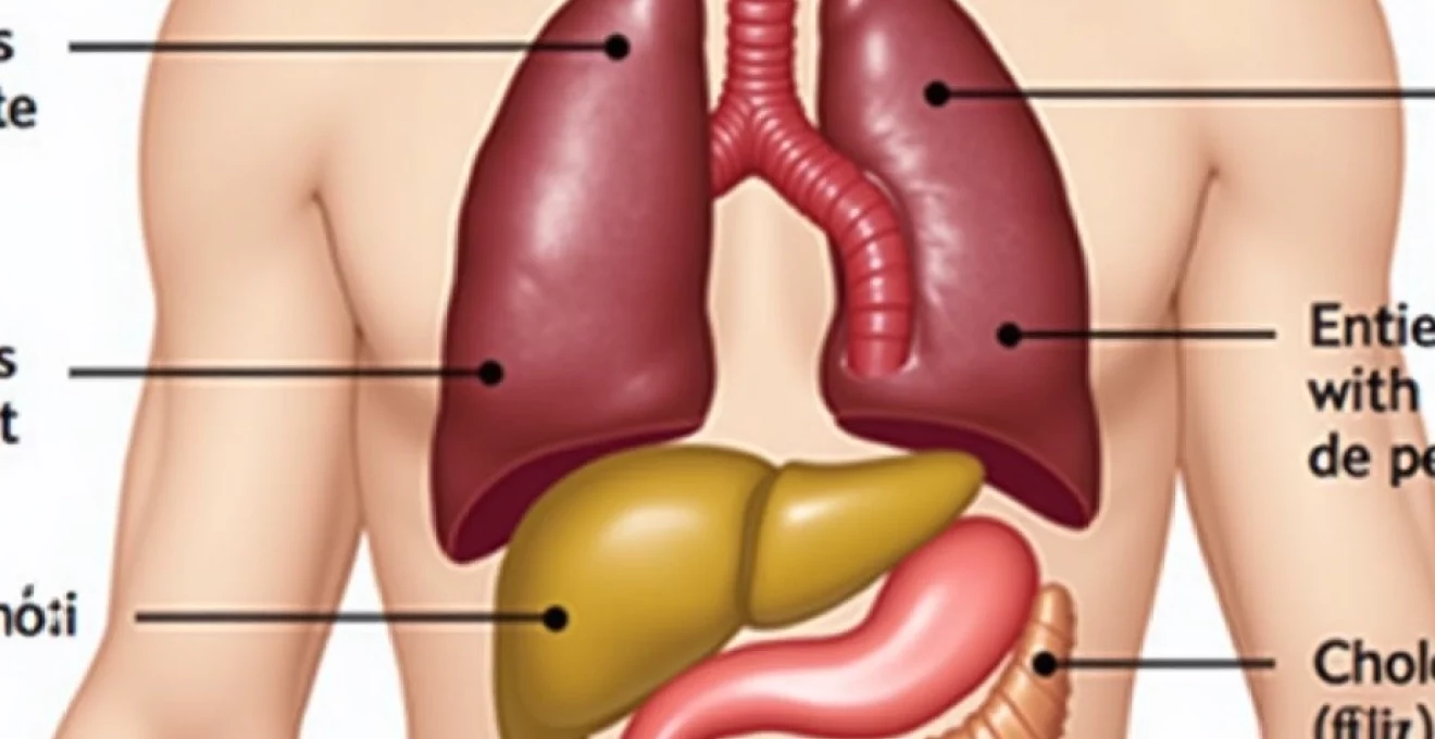
The right subcostal region encompasses the anatomical space immediately beneath the lower border of the right costal margin, extending from the midline to the posterior axillary line. This area contains a complex arrangement of visceral organs, muscular structures, and neural pathways that contribute to the varied presentations of pain experienced in this region. The overlapping innervation patterns create a phenomenon where pain originating from one structure may be perceived in seemingly unrelated anatomical locations.
Intercostal nerve distribution and right hypochondrium innervation
The intercostal nerves T6 through T12 provide the primary sensory innervation to the right hypochondrium and subcostal region. These nerves emerge from the thoracic spinal cord and traverse the intercostal spaces, sending branches that innervate both the overlying musculature and the underlying visceral structures. The T7-T9 nerve roots specifically contribute to hepatic innervation, whilst T10-T11 provide sensory input from the gallbladder and duodenum.
The complexity of this neural network explains why patients often describe pain that seems to radiate or shift between different areas of the right upper quadrant. Sympathetic nerve fibres from the celiac plexus also contribute to visceral pain transmission, particularly from hepatobiliary structures. This dual innervation pattern means that inflammatory or mechanical irritation of organs within the right subcostal region can produce pain sensations that vary significantly in character, intensity, and distribution.
Visceral pain versus somatic pain differentiation in right upper quadrant
Understanding the distinction between visceral and somatic pain patterns proves crucial for accurate diagnosis of right subcostal discomfort. Visceral pain typically presents as a deep, poorly localised aching sensation that patients often describe as cramping or pressure-like. This type of pain originates from the internal organs and tends to be accompanied by autonomic symptoms such as nausea, sweating, or changes in heart rate.
Conversely, somatic pain arising from the parietal peritoneum, muscle, or skeletal structures presents as sharp, well-localised discomfort that patients can often point to with precision. The character of somatic pain tends to be more constant and may worsen with movement or palpation. This distinction becomes particularly important when evaluating patients with suspected gallbladder disease , as the progression from visceral biliary colic to somatic peritoneal irritation often indicates the development of complications such as cholecystitis or perforation.
Referred pain patterns from T6-T12 dermatomes
The phenomenon of referred pain plays a significant role in the presentation of right subcostal discomfort, with pain originating from deep visceral structures being perceived in cutaneous areas sharing similar embryological origins or neural pathways. Classic examples include hepatic pain being felt in the right shoulder tip via the phrenic nerve (C3-C5), and gallbladder pain radiating to the right scapular region through T6-T9 dermatomes.
These referred pain patterns can sometimes mislead both patients and healthcare providers, particularly when the referred component predominates over the primary site of pathology. Understanding these neuroanatomical relationships allows clinicians to recognise that shoulder tip pain in the context of right subcostal discomfort may indicate diaphragmatic irritation secondary to hepatic inflammation or biliary pathology.
Costal margin anatomy and rib cage biomechanics
The anatomical structure of the costal margin itself can contribute to pain generation, particularly in conditions affecting the lower ribs or costal cartilages. The 8th, 9th, and 10th ribs, known as false ribs, connect to the sternum indirectly through costal cartilages, whilst the 11th and 12th ribs remain unattached anteriorly. This anatomical arrangement creates areas of relative weakness that may be susceptible to injury or inflammation.
Costochondritis, representing inflammation of the costal cartilages, frequently presents as sharp, localised pain that worsens with respiratory movements or chest wall palpation. The biomechanical forces acting on the rib cage during breathing, coughing, or physical activity can exacerbate pain arising from these structures, creating a distinctive pattern of symptoms that differs markedly from visceral pain sources.
Hepatobiliary disorders causing right subcostal sharp pain
The hepatobiliary system represents the most common source of pathology leading to right subcostal pain, with gallstone-related disorders accounting for over 80% of cases requiring surgical intervention. The intricate relationship between the liver, gallbladder, and biliary tree means that dysfunction in one component often affects the entire system, creating complex patterns of pain and associated symptoms that require careful evaluation and management.
Acute cholecystitis and Gallstone-Related biliary colic episodes
Acute cholecystitis develops when gallstones obstruct the cystic artery or when inflammation occurs within the gallbladder wall itself. The resulting pain typically begins as episodic biliary colic but progresses to constant, severe right subcostal pain that may radiate to the right shoulder or scapular region. This progression reflects the transition from intermittent ductal obstruction to sustained inflammatory changes within the gallbladder wall.
The classic presentation includes Murphy’s sign, where inspiration is interrupted by pain when the examiner palpates the right subcostal region during deep inspiration. Recent studies indicate that approximately 10-20% of adults in developed countries harbour gallstones , though only a fraction develop symptomatic disease. The risk factors for gallstone formation include female gender, obesity, rapid weight loss, and certain ethnic backgrounds, with the “4 F’s” (female, forty, fat, fertile) serving as a traditional mnemonic.
Biliary colic episodes typically last between 15 minutes and 5 hours, with pain intensity often described as severe enough to cause patients to seek emergency medical care. The pain may be accompanied by nausea, vomiting, and low-grade fever, particularly when inflammation progresses to acute cholecystitis. Laboratory investigations often reveal elevated white blood cell counts and inflammatory markers, whilst imaging studies such as ultrasound or HIDA scans can confirm the diagnosis.
Hepatomegaly secondary to fatty liver disease and hepatitis
Hepatomegaly, or liver enlargement, commonly presents as right subcostal fullness or discomfort rather than acute sharp pain. However, rapid hepatic enlargement due to acute hepatitis or severe fatty infiltration can stretch the hepatic capsule, producing sharp, constant pain that may worsen with movement or deep inspiration. Non-alcoholic fatty liver disease (NAFLD) has become increasingly prevalent, affecting up to 30% of adults in Western populations.
The pain associated with hepatic enlargement typically presents as a dull ache that may occasionally become sharp, particularly during periods of rapid expansion or when inflammatory changes affect the hepatic capsule. Patients often describe a sensation of fullness or pressure rather than the severe, cramping pain characteristic of biliary colic. Associated symptoms may include fatigue, nausea, and in cases of viral hepatitis, systemic symptoms such as fever and malaise.
Choledocholithiasis and common bile duct obstruction
Choledocholithiasis, the presence of stones within the common bile duct, represents a potentially serious complication of gallstone disease that can lead to biliary obstruction, cholangitis, or pancreatitis. The pain associated with common bile duct stones often differs from simple gallbladder colic, presenting as more persistent discomfort that may be accompanied by jaundice and dark urine.
The clinical presentation typically includes the classic Charcot’s triad of fever, jaundice, and right upper quadrant pain, though this complete constellation occurs in only approximately 50-75% of cases.
The development of jaundice in conjunction with right subcostal pain should always prompt immediate evaluation for biliary obstruction, as delayed treatment can lead to severe complications including cholangitis and hepatic dysfunction.
Cholangitis and ascending biliary tract infections
Ascending cholangitis represents a medical emergency characterised by bacterial infection of the biliary tree, typically occurring in the setting of biliary obstruction. The condition presents with severe right subcostal pain, high fever with rigors, and often progresses rapidly to sepsis if not promptly recognised and treated. The pain pattern may differ from simple biliary colic, often being more constant and severe.
Reynolds’ pentad, which includes the classic Charcot’s triad plus shock and altered mental status, indicates severe cholangitis with systemic complications. Mortality rates for untreated cholangitis can exceed 50% , emphasising the critical importance of early recognition and intervention. Treatment typically requires urgent biliary drainage through endoscopic or percutaneous approaches, combined with broad-spectrum antibiotic therapy.
Gastrointestinal pathology in right upper quadrant pain syndromes
Beyond hepatobiliary disorders, various gastrointestinal conditions can manifest as sharp right subcostal pain. These conditions often present diagnostic challenges due to overlapping symptoms and shared neural pathways, requiring careful clinical assessment to differentiate between different pathological processes affecting the digestive system.
Duodenal ulcer perforation and peptic disease complications
Duodenal ulcers, whilst classically associated with epigastric pain, can present with right subcostal discomfort, particularly when complicated by perforation or penetration into adjacent structures. The pain from duodenal ulcer disease typically follows a pattern related to meal timing, with symptoms often improving with food intake but worsening 2-3 hours postprandially when gastric acid production peaks.
Perforated duodenal ulcers represent surgical emergencies that can present with sudden-onset, severe right upper quadrant pain as gastric contents irritate the peritoneum. The pain may initially be localised to the epigastrium before spreading to involve the entire abdomen. The incidence of peptic ulcer disease has declined significantly following the recognition of Helicobacter pylori as a primary causative factor and the development of effective eradication therapies.
Hepatic flexure syndrome and colonic gas entrapment
Hepatic flexure syndrome, also known as right colic flexure syndrome, results from gas accumulation or spasm in the hepatic flexure of the colon. This condition can produce sharp, cramping pain in the right subcostal region that may be mistaken for more serious pathology. The pain typically occurs after meals and may be relieved by changes in position or passage of flatus.
The anatomical location of the hepatic flexure, positioned beneath the liver and adjacent to the gallbladder, means that pain from colonic distension can be difficult to distinguish from hepatobiliary pathology based on location alone. However, the episodic nature of the pain and its relationship to bowel function often provide diagnostic clues. Dietary modifications and increased physical activity frequently provide effective symptom relief for this benign condition.
Gastroesophageal reflux disease with Right-Sided epigastric extension
Gastroesophageal reflux disease (GERD) can occasionally present with pain that extends into the right subcostal region, particularly when associated with severe esophagitis or Barrett’s esophagus. The pain typically has a burning quality and may be accompanied by classic reflux symptoms such as heartburn, regurgitation, and nocturnal cough.
Atypical presentations of GERD may include chest pain that radiates to the right upper quadrant, potentially mimicking cardiac or biliary pathology. The relationship between symptoms and meal timing, position, and response to acid-suppressing medications often helps establish the diagnosis.
Up to 20% of adults in Western countries experience weekly symptoms of gastroesophageal reflux, making it one of the most prevalent gastrointestinal disorders.
Pyloric stenosis and gastric outlet obstruction
Pyloric stenosis in adults, whilst rare compared to the infantile form, can present with right subcostal pain associated with postprandial vomiting and early satiety. The condition may result from peptic ulcer disease, malignancy, or inflammatory conditions affecting the pyloric channel. The pain typically worsens after eating as gastric distension increases against the obstructed outlet.
Gastric outlet obstruction from any cause can produce a characteristic pattern of symptoms including projectile vomiting of undigested food, progressive weight loss, and electrolyte abnormalities. The pain pattern often includes both epigastric and right subcostal components due to gastric distension and secondary effects on adjacent structures.
Musculoskeletal aetiologies of sharp intercostal pain
Musculoskeletal causes of right subcostal pain are frequently overlooked in the differential diagnosis, yet they represent a significant proportion of cases presenting to primary care providers. These conditions often result from trauma, overuse, or inflammatory processes affecting the chest wall structures, and typically present with characteristic patterns that help distinguish them from visceral pathology.
Costochondritis, inflammation of the costal cartilages connecting the ribs to the sternum, commonly affects the lower ribs and can produce sharp, stabbing pain that worsens with respiratory movements, coughing, or chest wall palpation. The pain is typically well-localised and may be reproduced by applying pressure to specific points along the costal margin. Unlike visceral pain, costochondritis rarely produces associated systemic symptoms such as nausea or fever.
Intercostal muscle strain represents another common cause of right subcostal pain, particularly in individuals engaged in activities involving repetitive twisting motions or heavy lifting. The pain from muscle strain typically has an aching quality that may become sharp with specific movements or positions. Physical examination reveals tenderness along the affected muscle groups, and pain may be reproduced by resisted movements that engage the intercostal muscles.
Rib fractures, whether from direct trauma or pathological causes such as osteoporosis, produce characteristic sharp pain that intensifies with inspiration, coughing, or movement. The pain from rib fractures is typically severe and localised to the fracture site, with patients often adopting shallow breathing patterns to minimise discomfort. Complications from rib fractures can include pneumothorax or injury to underlying organs, particularly with fractures of the lower ribs that may damage the liver or spleen.
Slipping rib syndrome, a condition where the 8th, 9th, or 10th ribs become hypermobile due to weakness in the connecting fibrous tissue, can produce intermittent sharp pain that may be accompanied by a clicking or popping sensation. This condition is often triggered by sudden movements or specific positions and may be diagnosed using the “hooking maneuver” test during physical examination.
Pulmonary conditions manifesting as Right-Sided subcostal discomfort
Respiratory pathology can produce right subcostal pain through several mechanisms, including pleural irritation, diaphragmatic involvement, or referral patterns from lower lobe pneumonia. The proximity of the right lung base to the diaphragm and upper
abdominal organs creates multiple pathways through which pulmonary conditions can manifest as right subcostal pain. Lower lobe pneumonia, particularly when affecting the right lung base, commonly produces referred pain to the right subcostal region due to shared innervation patterns between the phrenic nerve and intercostal nerves.
Pleuritis or pleurisy affecting the right lower pleural space can produce sharp, stabbing pain that characteristically worsens with inspiration and coughing. The pain from pleural inflammation tends to be well-localised and may be accompanied by a distinctive pleural friction rub audible on auscultation. Patients often describe the sensation as feeling like someone is stabbing them with each breath , and they may adopt shallow breathing patterns to minimise discomfort.
Pneumothorax, particularly when involving the right lung, can present with sudden-onset sharp pain in the right subcostal region, accompanied by dyspnoea and reduced breath sounds on the affected side. Small pneumothoraces may produce localised pain without significant respiratory compromise, whilst larger pneumothoraces can rapidly progress to life-threatening tension pneumothorax requiring emergency decompression.
Pulmonary embolism represents a potentially life-threatening condition that may present with right-sided chest pain extending into the subcostal region, particularly when involving the lower lobe pulmonary vessels. The pain is typically sudden in onset and may be accompanied by dyspnoea, tachycardia, and in severe cases, haemoptysis. Risk factors include recent immobilisation, surgery, malignancy, or thrombophilic disorders.
The diagnosis of pulmonary embolism requires high clinical suspicion, as up to 30% of cases may present with atypical symptoms that can mimic other conditions causing right subcostal pain.
Diaphragmatic irritation from various causes, including subphrenic abscesses or hepatic pathology, can produce sharp pain that may be perceived in the right subcostal region. The diaphragm receives dual innervation from both the phrenic nerve (C3-C5) and intercostal nerves (T6-T12), explaining why diaphragmatic irritation can produce pain patterns that extend from the shoulder to the lower chest wall.
Emergency red flag symptoms requiring immediate medical intervention
Recognition of emergency presentations involving right subcostal pain is crucial for preventing life-threatening complications and ensuring appropriate urgent management. Certain symptom combinations and clinical presentations warrant immediate medical evaluation and should never be ignored or dismissed as benign conditions. Healthcare providers and patients alike must maintain awareness of these critical warning signs.
The presence of jaundice in conjunction with right subcostal pain represents a significant red flag that may indicate biliary obstruction, cholangitis, or severe hepatic dysfunction. Progressive jaundice accompanied by dark urine and pale stools suggests complete or near-complete biliary obstruction requiring urgent intervention to prevent irreversible hepatic damage. The development of altered mental status or signs of sepsis in this context indicates cholangitis with systemic complications, representing a true medical emergency.
Severe, unrelenting pain that fails to respond to appropriate analgesia may indicate complications such as gallbladder perforation, bowel obstruction, or vascular emergencies. The character of the pain often provides important clues, with sudden-onset, tearing sensations potentially indicating vascular pathology, whilst progressive, worsening pain may suggest inflammatory processes or mechanical obstruction.
Haemodynamic instability, including hypotension, tachycardia, or signs of shock, in the context of right subcostal pain demands immediate evaluation for conditions such as ruptured hepatic lesions, massive pulmonary embolism, or sepsis secondary to biliary or hepatic infections. The combination of abdominal pain with cardiovascular collapse should always prompt consideration of intra-abdominal bleeding or systemic infection.
Respiratory compromise accompanying right subcostal pain may indicate tension pneumothorax, massive pulmonary embolism, or severe pleural effusion. Patients presenting with dyspnoea, reduced oxygen saturation, or asymmetrical chest expansion require immediate assessment and potential emergency interventions such as chest decompression or mechanical ventilation.
The presence of peritoneal signs, including guarding, rigidity, or rebound tenderness, suggests peritoneal irritation that may result from perforation of hollow organs, biliary peritonitis, or other causes of acute abdomen. The development of a rigid, board-like abdomen represents a surgical emergency requiring immediate operative intervention to prevent sepsis and death.
Neurological symptoms occurring alongside right subcostal pain, such as confusion, altered consciousness, or focal neurological deficits, may indicate hepatic encephalopathy, cerebrovascular events, or systemic toxicity. These presentations require comprehensive evaluation to identify and address both the neurological and abdominal components of the clinical syndrome.
Temperature extremes, including high fever with rigors or hypothermia, in the context of right subcostal pain often indicate serious infectious processes requiring urgent antibiotic therapy and source control. The combination of fever, jaundice, and right upper quadrant pain should immediately raise suspicion for cholangitis, whilst hypothermia may suggest overwhelming sepsis with poor prognosis if not promptly treated.
Studies demonstrate that delayed recognition of emergency presentations involving right subcostal pain can increase mortality rates by up to 300%, emphasising the critical importance of early identification and intervention.
Gastrointestinal bleeding, manifested by haematemesis, melaena, or haematochezia, combined with right subcostal pain may indicate complications of peptic ulcer disease, varices secondary to portal hypertension, or other causes of upper gastrointestinal bleeding. The combination requires urgent endoscopic evaluation and aggressive resuscitation to prevent exsanguination.
Pregnancy-related presentations involving right subcostal pain require special consideration, as conditions such as HELLP syndrome (Haemolysis, Elevated Liver enzymes, Low Platelets) or severe preeclampsia can rapidly progress to life-threatening complications for both mother and foetus. The combination of right upper quadrant pain, hypertension, and proteinuria in pregnancy demands immediate obstetric consultation and consideration for urgent delivery.
When evaluating patients with right subcostal pain, healthcare providers must maintain a systematic approach that includes assessment of vital signs, detailed pain characterisation, associated symptoms, and appropriate diagnostic studies. The key to successful management lies in rapid recognition of emergency presentations whilst avoiding unnecessary interventions for benign conditions. Patient education regarding warning signs and when to seek immediate medical attention remains an essential component of comprehensive care for individuals experiencing right subcostal pain.