Lamotrigine-induced cutaneous reactions represent one of the most clinically significant dermatological concerns associated with anticonvulsant therapy. As a widely prescribed medication for epilepsy and bipolar disorder, Lamictal affects approximately 10% of patients with various degrees of skin manifestations. These reactions range from benign maculopapular eruptions to life-threatening conditions such as Stevens-Johnson syndrome and toxic epidermal necrolysis. Understanding the visual characteristics and clinical progression of these rashes is crucial for both healthcare professionals and patients to ensure appropriate management and prevent potentially fatal outcomes.
The appearance of lamotrigine-associated rashes varies significantly depending on their underlying pathophysiology and severity. Early recognition becomes paramount when considering that severe cutaneous adverse reactions can develop rapidly, sometimes within days of initial presentation. The challenge lies in distinguishing between harmless hypersensitivity reactions and those requiring immediate medical intervention, as initial presentations may appear deceptively similar.
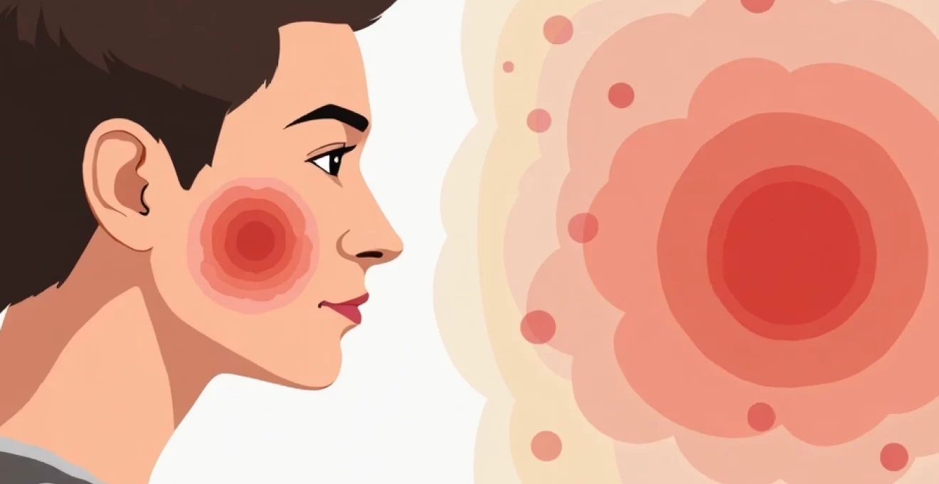
Lamictal-induced Stevens-Johnson syndrome: recognising early dermatological manifestations
Stevens-Johnson syndrome represents the most feared complication of lamotrigine therapy, occurring in approximately 0.08 to 1.3% of patients. This severe cutaneous adverse reaction typically manifests within the first eight weeks of treatment initiation, though cases have been documented outside this timeframe. The syndrome’s progression follows a characteristic pattern that begins subtly before rapidly evolving into a medical emergency requiring immediate hospitalisation.
Erythematous macular eruption: initial presentation patterns
The earliest manifestation of Stevens-Johnson syndrome often presents as discrete, irregularly shaped red macules measuring 1-3 centimetres in diameter. These lesions typically appear first on the trunk and face, displaying a characteristic target-like appearance with central pallor or dusky discolouration surrounded by erythematous borders. Unlike benign drug eruptions, these lesions demonstrate asymmetrical distribution and show a tendency to coalesce into larger, more extensive patches within 24-48 hours of onset.
The erythematous macules associated with Stevens-Johnson syndrome exhibit a distinctive morphology that differentiates them from common viral exanthems. They often display irregular borders with varying degrees of central necrosis, creating a mottled appearance that experienced clinicians recognise as highly concerning. The lesions typically feel warm to touch and may demonstrate slight elevation, progressing from flat macules to more palpable plaques as the condition advances.
Mucosal involvement: oral and conjunctival warning signs
One of the pathognomonic features of Stevens-Johnson syndrome is early mucosal involvement, which occurs in virtually all cases and often precedes cutaneous manifestations. The oral cavity typically shows the first signs, with painful erosions and ulcerations appearing on the lips, buccal mucosa, and tongue. These lesions begin as small vesicles or bullae that quickly rupture, leaving behind shallow, extremely painful ulcers with irregular borders and greyish-white bases.
Conjunctival involvement presents as bilateral erythema, oedema, and subsequent erosion of the palpebral and bulbar conjunctivae. Patients frequently report severe photophobia, excessive tearing, and a burning sensation that progressively worsens. The presence of pseudomembranes or frank membrane formation across the conjunctival surface indicates severe disease and heightens the risk of long-term ocular complications, including symblepharon formation and corneal scarring.
Nikolsky sign: epidermal detachment assessment techniques
The Nikolsky sign serves as a critical diagnostic indicator for severe cutaneous adverse reactions, including Stevens-Johnson syndrome. This clinical test involves applying gentle lateral pressure to apparently normal skin adjacent to lesional areas. A positive result, characterised by superficial epidermal sloughing or blister formation under minimal pressure, indicates widespread keratinocyte necrosis and epidermal-dermal junction compromise.
Healthcare providers perform this assessment by placing a finger on normal-appearing skin near the rash border and applying gentle sliding pressure. The immediate separation of the epidermis from the dermis signals extensive tissue damage and helps differentiate Stevens-Johnson syndrome from less severe conditions. This sign typically becomes positive as the syndrome progresses beyond its initial stages, making it a valuable tool for severity assessment and prognosis determination.
Fever and systemic symptoms: constitutional warning indicators
Constitutional symptoms accompanying lamotrigine-induced Stevens-Johnson syndrome often precede cutaneous manifestations by 1-3 days, creating an important window for early recognition. High-grade fever, typically exceeding 38.5°C, represents one of the most consistent early warning signs. This fever pattern differs from typical viral illnesses by its persistent nature and poor response to standard antipyretic medications.
Associated systemic symptoms include severe malaise, myalgia, arthralgia, and headache, creating a flu-like prodrome that may initially mislead both patients and healthcare providers. Gastrointestinal symptoms, including nausea, vomiting, and diarrhoea, frequently accompany the fever, while respiratory symptoms such as cough and dyspnoea may indicate pulmonary involvement. The combination of these constitutional symptoms with any new skin changes in patients taking lamotrigine should trigger immediate medical evaluation.
Benign lamotrigine hypersensitivity reactions: distinguishing mild cutaneous responses
Benign hypersensitivity reactions to lamotrigine encompass the majority of cutaneous adverse events, affecting approximately 8-9% of patients receiving the medication. These reactions, while concerning to patients, typically resolve spontaneously following dose reduction or medication discontinuation without long-term sequelae. Understanding their characteristic appearance helps differentiate them from more serious conditions and guides appropriate management decisions.
Morbilliform exanthem: generalised maculopapular distribution
The most common presentation of benign lamotrigine hypersensitivity manifests as a generalised morbilliform or measles-like eruption characterised by small, discrete, erythematous macules and papules. These lesions typically measure 2-5 millimetres in diameter and appear first on the trunk before spreading centrifugally to involve the extremities. The distribution pattern follows a symmetrical configuration, distinguishing it from the asymmetrical presentation seen in severe reactions.
The individual lesions maintain their discrete character without showing significant confluence or central changes. They appear bright red initially, gradually fading to a duller pink coloration over several days. Unlike Stevens-Johnson syndrome, these eruptions lack target-like morphology, mucosal involvement, or associated constitutional symptoms. Patients may experience mild pruritus, but severe pain or burning sensations are notably absent.
Anticonvulsant hypersensitivity syndrome: DRESS-Related manifestations
Drug Reaction with Eosinophilia and Systemic Symptoms (DRESS) syndrome represents a distinct entity within the spectrum of lamotrigine hypersensitivity reactions. This condition typically presents 2-8 weeks after treatment initiation with a characteristic triad of extensive skin eruption, fever, and internal organ involvement. The cutaneous manifestations often begin as a morbilliform eruption that progressively becomes more confluent and may develop into exfoliative dermatitis.
DRESS-associated rashes demonstrate several distinctive features that aid in diagnosis. The eruption typically shows facial oedema, particularly involving the periorbital regions, creating a characteristic appearance. Lymphadenopathy frequently accompanies the skin changes, with multiple enlarged lymph nodes palpable in cervical, axillary, and inguinal regions. Laboratory investigations reveal elevated eosinophil counts, often exceeding 1,500 cells per microlitre, along with elevated liver enzymes indicating hepatic involvement.
Urticarial reactions: wheals and angioedema presentation
Urticarial reactions to lamotrigine present as raised, erythematous wheals with well-defined borders and central pallor. These lesions vary significantly in size, ranging from small punctate wheals to large, geographic plaques covering extensive body surface areas. The characteristic feature of urticaria involves its transient nature, with individual lesions typically resolving within 24 hours while new ones may appear in different locations.
Accompanying angioedema may affect the face, lips, eyelids, and occasionally the larynx, creating potential airway concerns. The oedema appears asymmetrical and non-pitting, affecting deeper dermal and subcutaneous tissues. While urticarial reactions generally indicate a more benign hypersensitivity response, the presence of angioedema, particularly involving the upper airway, requires immediate medical attention and medication discontinuation.
Photosensitivity dermatitis: Sun-Exposed area involvement
Lamotrigine-induced photosensitivity manifests as erythematous, oedematous patches confined primarily to sun-exposed areas including the face, neck, dorsal hands, and forearms. This reaction pattern creates a distinctive distribution that clearly demarcates exposed from protected skin areas, often creating sharp cut-off lines at clothing margins. The severity varies from mild erythema resembling sunburn to more severe reactions with vesicle formation and subsequent desquamation.
Patients experiencing photosensitivity reactions typically report burning or stinging sensations in affected areas following sun exposure, with symptoms developing within hours of ultraviolet radiation contact. The reaction may persist for days to weeks after the initial exposure, gradually resolving with proper sun protection and, in some cases, requiring lamotrigine discontinuation to prevent recurrence.
Temporal progression: lamictal rash development timeline and risk factors
The temporal relationship between lamotrigine initiation and rash development follows predictable patterns that provide crucial diagnostic and prognostic information. Most cutaneous reactions manifest within the first eight weeks of treatment, with the highest risk period occurring between weeks 2-5. This timeline corresponds to the typical dose escalation phase, suggesting a strong correlation between dosing patterns and reaction probability.
Several factors significantly influence the likelihood and severity of lamotrigine-induced rashes. Rapid dose escalation represents the most modifiable risk factor, with studies demonstrating substantially higher reaction rates when recommended titration schedules are not followed. Concomitant valproate therapy doubles the risk of severe reactions by inhibiting lamotrigine metabolism, leading to elevated drug concentrations and prolonged exposure.
Patient age plays a critical role in determining reaction risk and severity. Paediatric patients demonstrate significantly higher rates of severe cutaneous adverse reactions, with children under 16 years showing a 1 in 300 risk of developing Stevens-Johnson syndrome compared to 1 in 1000 for adults. This age-related difference likely reflects developmental variations in drug metabolism and immune system responsiveness.
The critical importance of adhering to established titration schedules cannot be overstated, as rapid dose increases represent the single most preventable risk factor for severe cutaneous reactions.
Previous adverse reactions to other aromatic anticonvulsants, including phenytoin, carbamazepine, and phenobarbital, significantly increase the risk of cross-reactivity with lamotrigine. This phenomenon, known as anticonvulsant hypersensitivity syndrome, suggests shared immunological mechanisms across structurally similar compounds. Patients with a history of severe drug reactions require particularly careful monitoring and often benefit from alternative therapeutic approaches.
Paediatric versus adult lamotrigine cutaneous reactions: Age-Specific presentation differences
Age-related differences in lamotrigine-induced cutaneous reactions extend beyond simple risk stratification to encompass distinct presentation patterns and clinical courses. Paediatric patients typically develop more extensive and severe reactions, with rashes often covering larger body surface areas and showing greater tendency toward progression to serious conditions. The morphology of paediatric rashes may appear more dramatic, with brighter erythema and more pronounced oedema compared to adult presentations.
Adult lamotrigine rashes tend to follow more predictable patterns with clearer diagnostic features. The discrete nature of individual lesions remains more apparent in adults, facilitating easier differentiation between benign and serious reactions. Adults also demonstrate better correlation between symptom severity and ultimate outcomes, whereas children may develop serious complications from apparently mild initial presentations.
The speed of progression differs markedly between age groups, with paediatric cases showing more rapid evolution from initial skin changes to severe complications. This accelerated timeline necessitates more aggressive monitoring and lower thresholds for medication discontinuation in younger patients. Constitutional symptoms in children may be more subtle initially, making early recognition more challenging and emphasising the importance of maintaining high clinical suspicion.
Recovery patterns also vary significantly between adult and paediatric populations. Children often experience more prolonged healing phases with greater risk of post-inflammatory hyperpigmentation or scarring. However, they also demonstrate superior long-term recovery with less likelihood of permanent sequelae when appropriate treatment is instituted promptly. Adults typically show more predictable healing patterns but may require longer periods to achieve complete resolution.
Differential diagnosis: distinguishing lamictal rashes from viral exanthems and drug eruptions
Accurate differentiation between lamotrigine-induced rashes and other common cutaneous conditions requires systematic evaluation of morphological characteristics, distribution patterns, and associated symptoms. Viral exanthems represent the most common alternative diagnosis, particularly in paediatric patients, and share several overlapping features with drug-induced eruptions. However, careful attention to specific details usually reveals distinguishing characteristics that guide appropriate management decisions.
Viral exanthems typically demonstrate centripetal distribution patterns, beginning on the face and trunk before spreading to extremities, contrasting with the more random or centrifugal patterns seen in drug reactions. The temporal relationship to recent illness, presence of accompanying respiratory or gastrointestinal symptoms, and lack of correlation with medication timing help distinguish infectious causes. Additionally, viral rashes rarely involve mucosal surfaces in the dramatic fashion characteristic of severe drug reactions.
Other drug eruptions may share morphological similarities with lamotrigine reactions but often exhibit different temporal relationships and risk factor profiles. Antibiotic-induced rashes typically develop after several days of treatment rather than weeks, while NSAID reactions may show photo-distribution patterns distinct from lamotrigine photosensitivity. Careful medication history review, including over-the-counter preparations and supplements, helps identify alternative causative agents.
The key to accurate diagnosis lies in the systematic evaluation of morphology, distribution, timing, and associated symptoms rather than relying on any single characteristic feature.
Autoimmune conditions such as systemic lupus erythematosus or dermatomyositis may occasionally mimic drug-induced rashes, particularly when they present with photosensitive components. However, these conditions typically demonstrate additional systemic features, positive autoantibody profiles, and lack the clear temporal relationship to medication initiation seen in drug reactions. Laboratory investigations, including complement levels and autoantibody testing, may be necessary in unclear cases.
Contact dermatitis represents another important differential consideration, especially when rashes show localised distribution patterns. However, contact reactions typically demonstrate clear geometric patterns corresponding to allergen exposure sites and lack the widespread, symmetrical distribution characteristic of systemic drug reactions. Patch testing may be helpful in distinguishing topical allergen exposure from systemic drug hypersensitivity when the clinical picture remains unclear.
Emergency assessment protocols: when Lamotrigine-Induced rashes require immediate medical intervention
Establishing clear protocols for emergency assessment of lamotrigine-induced rashes proves essential for preventing serious morbidity and potential mortality. Healthcare providers must maintain high indices of suspicion for severe cutaneous adverse reactions while avoiding unnecessary anxiety and medication discontinuation for benign conditions. The challenge lies in creating assessment frameworks that capture serious cases early while minimising false-positive evaluations.
Immediate medical intervention becomes necessary when patients present with any combination of constitutional symptoms, mucosal involvement, or rapidly evolving skin changes. The presence of fever exceeding 38.5°C in conjunction with any new rash in lamotrigine-treated patients warrants emergency evaluation regardless of rash appearance. Similarly, any mucosal erosions or ulcerations, particularly involving the oral cavity or conjunctivae, indicate potential Stevens-Johnson syndrome development requiring urgent assessment.
Pain represents a critical distinguishing feature requiring immediate attention. While benign drug rashes may cause mild discomfort or pruritus, the presence of significant pain, burning, or stinging sensations suggests tissue necrosis and deeper injury. Patients reporting pain with skin contact, difficulty swallowing due to oral lesions, or severe photophobia with eye involvement require immediate hospital evaluation and probable admission for monitoring and treatment.
The percentage of body surface area involved provides another crucial assessment parameter. Rashes covering more than 30% of total body surface area, particularly when
showing rapid progression or associated with constitutional symptoms, requires urgent evaluation regardless of individual lesion characteristics.
Clinical photography documentation proves invaluable for tracking progression and facilitating specialist consultation when patients present with concerning rashes. Healthcare providers should photograph affected areas using standardised techniques, including overall distribution shots and close-up images of representative lesions. Serial photography allows objective assessment of progression rates and helps distinguish stable benign conditions from evolving serious reactions requiring intervention.
Laboratory investigations may provide supportive evidence for severe reactions but should not delay treatment decisions when clinical presentations warrant immediate action. Complete blood counts revealing eosinophilia, thrombocytopenia, or atypical lymphocytes suggest systemic involvement characteristic of DRESS syndrome or other severe hypersensitivity reactions. However, normal laboratory values do not exclude serious conditions, and clinical assessment remains paramount in emergency decision-making.
Emergency departments should maintain specific protocols for lamotrigine-related presentations, including immediate dermatology consultation availability and standardised assessment tools for severity grading. Staff training programs should emphasise recognition of early warning signs and the critical importance of medication history documentation. The development of decision-support tools incorporating risk stratification algorithms can help standardise care and reduce variability in management approaches across different healthcare settings.
Patient education regarding warning signs represents a crucial component of emergency prevention strategies. Individuals prescribed lamotrigine should receive clear written instructions describing concerning symptoms and specific situations requiring immediate medical attention. Educational materials should include visual references depicting various rash appearances and emphasise the importance of seeking prompt evaluation rather than waiting for symptoms to resolve spontaneously.
Early recognition and immediate intervention remain the cornerstones of preventing serious morbidity and mortality associated with severe lamotrigine-induced cutaneous reactions.
Healthcare systems must establish clear communication pathways between emergency departments, prescribing physicians, and specialist consultants to ensure coordinated care when serious reactions develop. This includes protocols for rapid medication discontinuation, alternative treatment initiation, and follow-up arrangements to monitor patient recovery and prevent recurrence. The implementation of standardised reporting systems helps track reaction patterns and contributes to ongoing safety surveillance efforts.
What warning signs should prompt patients to seek immediate medical care? Any combination of fever, mouth sores, eye irritation, or rapidly spreading rash requires urgent evaluation, as these symptoms may herald the development of life-threatening complications. The window for effective intervention narrows rapidly once severe reactions begin, making early recognition and prompt action essential for optimal outcomes.