Fingernail splitting down the middle represents one of the most frustrating nail conditions affecting millions of people worldwide. This phenomenon, medically termed onychorrhexis when occurring longitudinally, disrupts the nail’s structural integrity and can significantly impact both appearance and function. Understanding the complex interplay between nail anatomy, environmental factors, and underlying health conditions provides essential insight into prevention and treatment strategies. The nail plate’s sophisticated architecture relies on precise keratin formation and adequate nutritional support, making it vulnerable to various internal and external stressors that can compromise its strength and cohesion.
Nail plate anatomy and structural composition
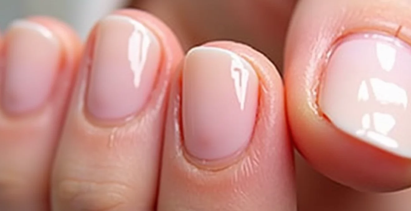
The fingernail represents a remarkable biological structure composed primarily of keratinised cells arranged in distinct layers. This complex architecture begins at the nail matrix, located beneath the proximal nail fold, where specialised cells continuously produce the proteins necessary for nail growth. The nail plate itself consists of approximately 25 layers of dead, flattened cells bound together by intercellular cement, creating a protective barrier that shields the sensitive nail bed beneath.
Keratin matrix architecture in fingernail formation
Keratin proteins form the fundamental building blocks of nail structure, with hard keratin comprising roughly 80-90% of the nail plate’s composition. These proteins organise into fibrous bundles that run parallel to the nail’s growth direction, creating natural ridges that contribute to the nail’s characteristic appearance. The keratin matrix contains both alpha-keratin filaments and keratin-associated proteins that crosslink to provide tensile strength. When this protein network becomes compromised through nutritional deficiencies or environmental damage, longitudinal splitting becomes increasingly likely.
Nail bed vascularisation and nutrient supply mechanisms
The nail bed’s rich vascular network supplies essential nutrients and oxygen to support healthy nail growth through a complex system of capillaries and arteriovenous anastomoses. This vascular supply delivers crucial minerals including zinc, iron, and selenium, which play vital roles in keratin synthesis and nail plate integrity. Compromised circulation, whether from systemic conditions or local trauma, can significantly impact nail quality and increase susceptibility to splitting. The nail bed’s unique anatomy also facilitates the removal of metabolic waste products that could otherwise accumulate and weaken the nail structure.
Cuticle and eponychium protective barrier functions
The cuticle and eponychium serve as critical protective barriers that prevent bacterial and fungal invasion while maintaining optimal moisture levels around the nail matrix. These structures produce natural oils and proteins that help seal the junction between the nail plate and surrounding skin tissue. When compromised through excessive manipulation or harsh chemical exposure, these protective barriers fail to maintain proper hydration balance, leading to increased brittleness and splitting tendencies. Professional nail technicians emphasise the importance of gentle cuticle care to preserve these natural defence mechanisms.
Longitudinal ridge formation and natural nail topography
Longitudinal ridges represent normal anatomical variations that become more pronounced with advancing age due to decreased cellular renewal rates in the nail matrix. These ridges follow the natural growth pattern of keratin fibres and can create weak points where splitting preferentially occurs. The depth and prominence of these ridges vary significantly between individuals and can be influenced by genetic factors, hormonal changes, and environmental exposures. Understanding this natural topography helps distinguish between normal age-related changes and pathological nail conditions requiring medical intervention.
Primary pathological causes of longitudinal nail splitting
Medical conditions affecting nail integrity encompass a broad spectrum of dermatological, systemic, and inflammatory disorders. These pathological processes can directly damage the nail matrix, alter keratin production, or compromise the structural proteins that maintain nail cohesion. Early recognition of these underlying causes enables targeted treatment approaches that address root causes rather than merely managing symptoms.
Onychorrhexis: brittle nail syndrome and keratin deficiency
Onychorrhexis represents a specific form of nail dystrophy characterised by longitudinal ridging and splitting that results from defective keratinisation processes. This condition frequently manifests as multiple parallel splits extending from the free edge toward the nail base, creating a characteristic “washboard” appearance. The underlying pathophysiology involves disrupted protein synthesis within the nail matrix, leading to weakened intercellular bonds and increased susceptibility to mechanical stress. Research indicates that approximately 20% of the adult population experiences some degree of nail brittleness, with women over 50 being disproportionately affected.
Trachyonychia and Twenty-Nail dystrophy manifestations
Trachyonychia, commonly known as twenty-nail dystrophy when affecting all fingernails and toenails, presents as rough, sandpaper-like nail surfaces with prominent longitudinal ridging. This condition often precedes or accompanies splitting episodes due to the compromised nail plate structure. The rough surface texture results from irregular keratinisation patterns within the nail matrix, creating areas of differential strength that predispose to fracture formation. Trachyonychia can be associated with various autoimmune conditions, including alopecia areata and vitiligo, suggesting shared pathogenic mechanisms involving immune-mediated matrix dysfunction.
Lichen Planus-Induced nail matrix scarring
Lichen planus affecting the nail apparatus can cause permanent matrix scarring that disrupts normal nail formation and increases splitting risk. This inflammatory condition creates chronic inflammation within the nail matrix, leading to fibrosis and altered keratin production patterns. The resulting nail changes often include longitudinal ridging, splitting, and in severe cases, complete nail loss. Early intervention with topical or systemic immunosuppressive agents can help prevent irreversible matrix damage and preserve nail function.
Psoriatic arthritis nail involvement and pitting patterns
Psoriatic nail disease affects up to 90% of individuals with psoriatic arthritis and frequently includes longitudinal splitting alongside characteristic nail pitting. The inflammatory process targets the nail matrix and bed simultaneously, creating focal areas of weakness where splits commonly initiate. These splits often occur in conjunction with oil drop discolouration and subungual hyperkeratosis, creating a distinctive clinical presentation. The severity of nail involvement often correlates with joint disease activity, making nail examination an important component of rheumatological assessment.
Thyroid dysfunction impact on nail plate integrity
Both hyperthyroidism and hypothyroidism significantly affect nail growth rate and structural integrity through their influence on cellular metabolism and protein synthesis. Hypothyroidism commonly causes slow growth, brittleness, and increased splitting tendency due to reduced metabolic activity within the nail matrix. Conversely, hyperthyroidism can lead to rapid but structurally defective nail growth that predisposes to splitting. Thyroid hormone directly regulates keratinocyte proliferation and differentiation, making nail changes an early indicator of thyroid dysfunction that may precede other clinical symptoms.
Environmental and mechanical trauma factors
Environmental exposures and repetitive mechanical trauma represent the most common preventable causes of longitudinal nail splitting. These external factors can progressively weaken nail structure through protein denaturation, lipid extraction, and mechanical stress concentration at vulnerable points. Understanding these mechanisms enables the development of effective protective strategies that significantly reduce splitting risk in high-exposure individuals.
Repetitive microtrauma from occupational hand use
Occupational activities involving repetitive finger use, such as typing, musical instrument playing, or manual labour, create cumulative microtrauma that weakens nail structure over time. These activities generate repetitive stress cycles that can initiate microscopic fractures at the nail’s free edge, which then propagate proximally along natural ridge lines. Healthcare workers, food service employees, and individuals in cleaning industries face particularly high risk due to combined mechanical and chemical exposures. Protective strategies including appropriate glove selection and technique modification can significantly reduce occupational nail damage.
Chemical exposure effects from acetone and formaldehyde
Acetone-based nail polish removers and formaldehyde-containing nail hardeners represent significant chemical threats to nail integrity through their dehydrating and protein-denaturing effects. Acetone strips natural oils from the nail plate, reducing flexibility and increasing brittleness that predisposes to splitting. Formaldehyde creates crosslinks between keratin proteins that initially strengthen nails but ultimately lead to rigidity and fracture susceptibility. Regular exposure to these chemicals requires careful moisture replacement and protective barrier application to maintain nail health.
Excessive manicure cuticle manipulation damage
Aggressive cuticle manipulation during manicure procedures can damage the nail matrix and create inflammatory responses that compromise nail formation. Excessive pushing, cutting, or chemical removal of cuticles disrupts the protective seal around the nail base, allowing moisture loss and bacterial invasion. This trauma can extend into the matrix area, creating localised scarring that results in permanent nail deformities and increased splitting tendency. Professional nail technicians increasingly advocate for minimal cuticle manipulation to preserve nail health.
UV light degradation of nail keratin proteins
Ultraviolet radiation exposure causes photochemical damage to nail keratin through free radical formation and protein crosslinking disruption. This process weakens the nail’s structural integrity and creates areas of differential strength that predispose to splitting. Individuals with significant sun exposure or those using UV nail lamps for gel manicures face increased risk of photodamage-related nail problems. Recent studies suggest that regular UV exposure may accelerate age-related nail changes and increase splitting frequency in susceptible individuals.
Nutritional deficiencies and systemic health correlations
Nutritional status profoundly influences nail health through its impact on keratin synthesis, cellular metabolism, and structural protein formation. Deficiencies in specific vitamins, minerals, and amino acids can compromise nail integrity and increase splitting susceptibility. The nail plate serves as a useful indicator of nutritional status, with changes often appearing before other clinical signs of deficiency become apparent. Biotin deficiency represents one of the most well-documented nutritional causes of nail brittleness and splitting, with supplementation showing consistent benefits in clinical studies.
Essential fatty acid deficiencies particularly affect nail flexibility and moisture retention, as these lipids play crucial roles in maintaining intercellular cement integrity. Omega-3 fatty acids contribute to anti-inflammatory processes that protect against matrix damage, while omega-6 fatty acids support cellular membrane stability. Iron deficiency anaemia creates characteristic nail changes including koilonychia (spoon-shaped nails) and increased brittleness that predisposes to splitting. Protein malnutrition affects keratin synthesis directly, leading to structurally defective nails with reduced tensile strength.
Clinical studies demonstrate that biotin supplementation at doses of 2.5mg daily can improve nail thickness and reduce splitting by up to 25% within six months of consistent use.
Zinc deficiency impacts multiple aspects of nail formation, including matrix cell division, keratin synthesis, and immune function that protects against secondary infections. This mineral serves as a cofactor for over 300 enzymatic reactions involved in protein metabolism and cellular repair processes. Vitamin D deficiency increasingly recognised as contributing to nail problems through its effects on calcium homeostasis and cellular differentiation. Adequate vitamin D levels support proper keratinocyte maturation and strengthen the nail’s calcium-dependent structural proteins.
Professional diagnostic assessment and dermoscopy evaluation
Professional evaluation of longitudinal nail splitting requires systematic assessment using standardised diagnostic criteria and advanced imaging techniques. Dermoscopy provides detailed visualisation of nail plate architecture, revealing subtle changes that may indicate underlying pathology or guide treatment selection. Clinical evaluation begins with comprehensive medical history taking, focusing on onset patterns, associated symptoms, occupational exposures, and medication use. Physical examination includes assessment of all twenty nails, surrounding skin, and evaluation for systemic signs of underlying disease.
Dermoscopic examination reveals characteristic features that help differentiate between various causes of nail splitting, including ridge patterns, colour variations, and vascular changes within the nail bed. Advanced diagnostic techniques such as nail plate biopsy or matrix sampling may be indicated in cases where malignancy or inflammatory conditions are suspected. Laboratory investigations should include complete blood count, thyroid function tests, and nutritional assessments when systemic causes are considered. Mycological examination helps exclude fungal infections that can mimic or contribute to splitting nail conditions.
Professional nail assessment should always include evaluation of the contralateral nails and examination of family members when genetic conditions are suspected, as this information significantly influences treatment approaches.
Photographic documentation provides valuable baseline information for monitoring treatment response and identifying subtle progressive changes over time. Digital imaging with standardised lighting and positioning enables objective assessment of nail improvements following therapeutic interventions. Nail plate thickness measurements using ultrasound or optical coherence tomography offer quantitative data that supplements clinical observations and helps guide treatment intensity.
Evidence-based treatment protocols and prevention strategies
Contemporary treatment approaches for longitudinal nail splitting emphasise combination therapies that address both underlying causes and symptomatic relief. Evidence-based protocols incorporate topical therapies, systemic treatments, and preventive measures tailored to individual patient needs and specific causative factors. Topical treatments focus on restoring nail plate hydration, strengthening protein structure, and protecting against further environmental damage. Urea-containing preparations at concentrations of 10-40% demonstrate significant efficacy in improving nail flexibility and reducing splitting frequency.
Systemic interventions target nutritional deficiencies and underlying medical conditions that contribute to nail fragility. Biotin supplementation remains the most evidence-supported nutritional intervention, with multiple randomised controlled trials demonstrating significant improvements in nail quality and reduced splitting rates. Collagen peptide supplementation shows emerging promise for nail strengthening, with preliminary studies indicating improvements in nail growth rate and structural integrity. Treatment duration typically requires 3-6 months before significant clinical improvements become apparent due to the slow nail growth rate.
Professional nail hardening treatments using controlled formaldehyde exposure or alternative strengthening agents can provide temporary improvement while addressing underlying causes, but require careful monitoring to prevent excessive rigidity that may worsen splitting tendencies.
Preventive strategies form the cornerstone of long-term nail health maintenance and include both lifestyle modifications and protective measures. Regular application of nail and cuticle moisturisers helps maintain optimal hydration levels and prevents excessive drying that predisposes to splitting. Protective glove use during household chores, gardening, and chemical exposures significantly reduces environmental damage to nail structure. Gentle nail care practices, including proper filing techniques and avoiding aggressive cuticle manipulation, minimise mechanical trauma that can initiate splitting episodes.
Professional manicure techniques should emphasise nail health preservation over aggressive cosmetic procedures that may compromise structural integrity. Regular nail trimming using sharp, clean instruments helps prevent split propagation and reduces mechanical stress on weakened areas. Dietary optimisation focusing on adequate protein intake, essential fatty acids, and key micronutrients supports ongoing nail formation and repair processes. Long-term success requires patient education about nail anatomy, risk factors, and the importance of consistent preventive care practices in maintaining optimal nail health throughout life.