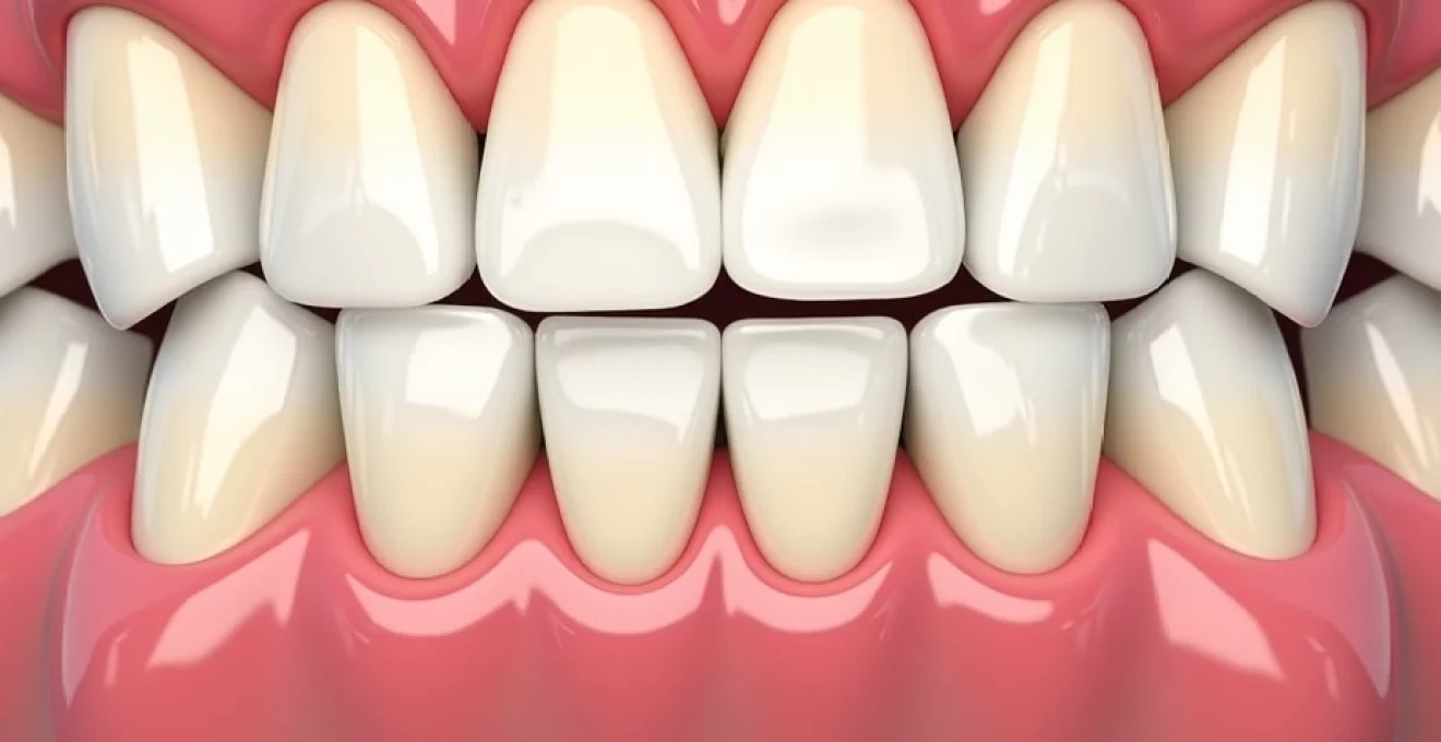
Lower front tooth pain affects millions of people worldwide and can significantly impact daily activities such as eating, speaking, and maintaining proper oral hygiene. The mandibular incisors, comprising the four front teeth in your lower jaw, are particularly susceptible to various dental conditions due to their prominent position and constant exposure to external stimuli. Understanding the underlying causes of this discomfort is crucial for seeking appropriate treatment and preventing further complications.
These teeth play a vital role in biting, cutting food, and supporting proper speech patterns. When pain develops in this region, it often signals underlying dental pathologies that require immediate attention. The complexity of lower anterior tooth pain stems from the intricate network of nerves, blood vessels, and supporting structures that can become compromised through various mechanisms.
Early intervention remains the most effective approach to managing lower front tooth pain, as delayed treatment can lead to more severe complications and extensive procedures. The multifactorial nature of mandibular incisor discomfort necessitates a comprehensive understanding of potential causes to ensure accurate diagnosis and appropriate treatment planning.
Common dental pathologies affecting lower anterior teeth
Several dental pathologies commonly affect the lower front teeth, each presenting with distinct characteristics and requiring specific treatment approaches. These conditions often develop gradually, making early detection challenging without regular dental examinations. The unique anatomy and position of mandibular incisors predispose them to particular types of dental problems that may not commonly affect other teeth in the mouth.
Periodontal disease and gingival recession in mandibular incisors
Periodontal disease represents one of the most prevalent causes of lower front tooth pain, affecting the supporting structures surrounding these teeth. The condition typically begins as gingivitis, characterised by inflammation of the gingival tissues, and can progress to periodontitis if left untreated. Bacterial accumulation along the gingival margin creates toxins that trigger inflammatory responses, leading to tissue breakdown and eventual bone loss.
Gingival recession commonly occurs in the lower anterior region due to aggressive brushing techniques, thin gingival biotype, and anatomical factors. When gums recede, the exposed root surfaces lack protective enamel coverage, making them highly sensitive to temperature changes, acidic foods, and mechanical stimulation. This exposure often results in sharp, shooting pain when consuming hot or cold beverages.
Advanced periodontal disease can lead to tooth mobility and eventual tooth loss if proper intervention is not implemented promptly.
Dental caries formation in lower central and lateral incisors
Dental caries in mandibular incisors often develop along the cervical margins where plaque accumulation is common. The lower front teeth are particularly vulnerable to decay due to their proximity to salivary gland openings, which can create areas of plaque stagnation. Acidogenic bacteria metabolise dietary sugars and starches, producing acids that demineralise tooth enamel and create cavities.
Interproximal caries between adjacent lower incisors can be particularly challenging to detect during routine visual examinations. These cavities often remain asymptomatic until they progress deeper into the tooth structure, reaching the dentine or pulp tissue. When decay advances to these levels, patients typically experience spontaneous pain, increased sensitivity, and discomfort when biting.
Enamel hypoplasia and developmental defects
Enamel hypoplasia affects the quality and quantity of enamel formation during tooth development, creating areas of weakness in the lower front teeth. These developmental defects can result from various factors including nutritional deficiencies, childhood illnesses, or genetic conditions. The compromised enamel structure makes these teeth more susceptible to decay, sensitivity, and structural failure.
Patients with enamel hypoplasia often report chronic sensitivity in their lower front teeth, particularly when exposed to acidic foods or temperature extremes. The irregular enamel surface also creates areas where plaque can accumulate more easily, increasing the risk of caries development and gingival inflammation.
Tooth sensitivity from exposed dentinal tubules
Dentinal hypersensitivity occurs when dentinal tubules become exposed through enamel loss or gingival recession, allowing external stimuli to reach the tooth’s nerve endings. This condition frequently affects the lower front teeth due to their thin enamel layer and susceptibility to erosion. Hydrodynamic theory explains how fluid movement within these tubules triggers nerve responses, resulting in sharp, transient pain.
Common triggers for dentinal sensitivity include cold air, sweet foods, acidic beverages, and mechanical stimulation during brushing or flossing. The pain typically subsides quickly once the stimulus is removed, distinguishing it from other types of dental pain that may persist longer.
Orthodontic and occlusal factors contributing to lower front tooth pain
Orthodontic and occlusal factors play a significant role in lower front tooth discomfort, often creating conditions that predispose these teeth to various problems. The relationship between tooth alignment, bite forces, and oral health is complex, with malocclusions potentially leading to uneven stress distribution and localised trauma.
Malocclusion-related trauma from overbite and overjet
Excessive overbite and overjet conditions can create traumatic occlusion patterns that subject the lower front teeth to abnormal forces during chewing and speaking. When upper teeth significantly overlap lower teeth, the mandibular incisors may experience constant pressure against the palatal tissues, leading to inflammation and discomfort. This chronic trauma can result in tooth mobility, pulpal inflammation, and eventual tooth loss if not corrected.
Deep bite conditions often cause the lower incisors to contact the palatal gingiva, creating ulcerations and chronic inflammation. Patients frequently report soreness in their lower front teeth, particularly after meals or extended periods of talking. The repetitive trauma can also lead to excessive wear patterns on the incisal edges of these teeth.
Bruxism and nocturnal grinding effects on mandibular incisors
Nocturnal bruxism subjects the lower front teeth to extraordinary forces that far exceed normal chewing pressures. During sleep, the protective reflexes that typically prevent excessive bite forces are diminished, allowing patients to generate pressures up to six times greater than normal. The mandibular incisors, being the first point of contact in many grinding patterns, bear significant stress during these episodes.
Chronic bruxism leads to characteristic wear patterns on the incisal edges of lower front teeth, often creating flat, abraded surfaces. Patients may wake with tooth pain, jaw stiffness, and increased sensitivity in their mandibular incisors. The constant grinding can also cause microfractures in the enamel, predisposing these teeth to caries and sensitivity.
Studies indicate that bruxism affects approximately 8-15% of the adult population, with many cases remaining undiagnosed until significant damage occurs.
Crowding-induced plaque accumulation and inflammation
Dental crowding in the lower anterior region creates areas that are difficult to clean effectively, leading to plaque accumulation and subsequent inflammation. When teeth are misaligned or overlapping, traditional brushing and flossing techniques may not adequately remove bacterial deposits from all surfaces. This plaque stagnation creates an environment conducive to caries development and periodontal disease.
The irregular tooth positioning associated with crowding also creates food traps where particles can become impacted, further contributing to bacterial growth and inflammation. Patients with crowded lower front teeth often experience localised gingival swelling, bleeding during oral hygiene procedures, and persistent discomfort in the affected areas.
Retainer-related pressure points and tissue irritation
Orthodontic retainers, while essential for maintaining tooth alignment, can sometimes create pressure points that cause discomfort in the lower front teeth. Fixed lingual retainers may irritate the tongue and gingival tissues, while removable retainers can create areas of concentrated pressure during insertion and removal. Poorly fitting retainers can exert excessive forces on individual teeth, leading to mobility and pain.
Patients wearing retainers may experience increased sensitivity in their lower front teeth, particularly during the initial adjustment period. Proper retainer maintenance and regular adjustments are crucial for preventing complications and ensuring patient comfort throughout the retention phase of orthodontic treatment.
Traumatic injuries and their sequelae in lower anterior dentition
Traumatic injuries to the lower front teeth can result from various incidents including sports activities, accidents, falls, and interpersonal violence. The prominent position of mandibular incisors makes them particularly vulnerable to direct trauma, which can cause immediate damage or lead to long-term complications that manifest as chronic pain. Understanding the types of traumatic injuries and their potential consequences is essential for proper diagnosis and treatment planning.
Dental trauma can be classified into several categories, including enamel fractures, dentine fractures, pulp exposures, root fractures, luxation injuries, and avulsion. Each type of injury presents unique challenges and may require different treatment approaches. Immediate assessment following trauma is crucial, as early intervention can often prevent more serious complications and preserve tooth vitality.
Crown fractures involving only enamel typically cause sharp edges that can irritate the tongue and lips, while also creating areas susceptible to plaque accumulation. More extensive fractures exposing dentine or pulp tissue require urgent treatment to prevent bacterial invasion and subsequent infection. Root fractures, though less common, can cause persistent pain and may necessitate complex treatment procedures or extraction.
Luxation injuries involve displacement of teeth from their normal position and can cause damage to the periodontal ligament and surrounding bone. These injuries often result in increased tooth mobility, sensitivity, and pain during function. The healing process following luxation injuries can be prolonged, with some teeth developing pulpal necrosis months or years after the initial trauma.
Post-traumatic complications may include pulpal necrosis, root resorption, ankylosis, and inflammatory resorption. These sequelae can develop gradually and may not become apparent until months or years following the original injury. Regular monitoring of traumatised teeth is essential to detect these complications early and implement appropriate interventions. The development of chronic pain in previously traumatised lower front teeth often indicates ongoing pathological processes that require professional evaluation and treatment.
Systemic conditions manifesting as lower front tooth discomfort
Various systemic conditions can manifest as pain or discomfort in the lower front teeth, highlighting the importance of considering the patient’s overall health status when evaluating dental complaints. These conditions may directly affect oral tissues or create referred pain patterns that localise to the mandibular anterior region.
Trigeminal neuralgia and mandibular nerve branch involvement
Trigeminal neuralgia represents a distinct neurological condition characterised by sudden, severe shooting pain along the distribution of the trigeminal nerve. When the mandibular division is affected, patients may experience intense pain that appears to originate from their lower front teeth. This neuropathic pain is typically triggered by light touch, speaking, chewing, or even gentle air movement across the face.
The pain associated with trigeminal neuralgia is often described as electric shock-like and can last from seconds to minutes. Unlike typical dental pain, this condition does not respond to conventional dental treatments and requires neurological intervention. Proper diagnosis is crucial to avoid unnecessary dental procedures and ensure appropriate medical management.
Temporomandibular joint disorders affecting anterior teeth
Temporomandibular joint disorders can create referred pain patterns that affect the lower front teeth, particularly when muscle spasm and joint dysfunction alter normal jaw mechanics. The complex relationship between TMJ function and dental occlusion means that joint problems can manifest as tooth pain, while dental problems can contribute to joint dysfunction.
Patients with TMJ disorders often report morning stiffness, clicking or popping sounds during jaw movement, and aching pain in their teeth and jaw muscles. The pain may be particularly noticeable in the lower front teeth due to their role in guiding jaw movements during function. Myofascial pain associated with TMJ disorders can create trigger points that refer pain to various areas of the head and neck, including the anterior mandibular region.
Vitamin deficiencies and Scurvy-Related gingival inflammation
Vitamin C deficiency, though rare in developed countries, can cause severe gingival inflammation and bleeding that may be mistaken for localised periodontal disease. Scurvy affects collagen synthesis, leading to weakened blood vessel walls and compromised healing capacity. The gingival tissues around the lower front teeth may become swollen, friable, and painful.
Other nutritional deficiencies, including B-complex vitamins and iron, can also manifest as oral symptoms that may cause discomfort in the lower anterior region. These deficiencies can compromise immune function and tissue healing, making patients more susceptible to opportunistic infections and delayed wound healing following dental procedures.
Hormonal fluctuations during pregnancy and menstruation
Hormonal changes during pregnancy and menstruation can significantly affect gingival health, often leading to increased inflammation and sensitivity in the lower front teeth. Elevated levels of oestrogen and progesterone increase vascular permeability and alter the immune response, making gingival tissues more susceptible to bacterial irritation. Pregnancy gingivitis commonly affects the anterior teeth due to their accessibility to plaque accumulation.
The hormonal influences on oral tissues can cause previously asymptomatic areas to become painful and inflamed. Women may notice increased bleeding during brushing and heightened sensitivity in their lower front teeth during these periods. Proper oral hygiene becomes even more crucial during times of hormonal fluctuation to prevent the development of more serious periodontal problems.
Pregnancy gingivitis affects up to 75% of pregnant women and typically begins in the second trimester when hormonal levels are highest.
Diagnostic techniques for evaluating lower incisor pain
Accurate diagnosis of lower front tooth pain requires a systematic approach combining clinical examination techniques, diagnostic imaging, and specialised tests to identify the underlying cause. The diagnostic process begins with a comprehensive history taking to understand the nature, duration, and characteristics of the patient’s symptoms. Clinicians must gather information about pain onset, triggers, relieving factors, and associated symptoms to narrow the differential diagnosis.
Clinical examination involves visual inspection, palpation, percussion testing, and mobility assessment of the affected teeth. Thermal testing using cold and heat stimuli helps evaluate pulpal vitality, while electric pulp testing can provide additional information about nerve function. Periodontal probing depths and bleeding indices assess the health of supporting structures around the lower front teeth.
Radiographic imaging plays a crucial role in diagnosing many conditions affecting the lower anterior teeth. Periapical radiographs provide detailed images of individual teeth and their surrounding structures, revealing caries, root fractures, periapical pathology, and bone loss. Bitewing radiographs are particularly useful for detecting interproximal caries between adjacent incisors that may not be visible clinically.
Advanced imaging techniques, including cone beam computed tomography, may be necessary for complex cases involving trauma, root resorption, or suspected pathology. These three-dimensional images provide superior visualisation of anatomical structures and can reveal details not apparent on conventional radiographs. Digital imaging also allows for image enhancement and measurement capabilities that aid in treatment planning.
Specialised diagnostic tests may include bite analysis, occlusal marking, and TMJ evaluation when occlusal factors are suspected. Sleep study referrals may be appropriate for patients with suspected bruxism, while medical consultations may be necessary when systemic conditions are considered. The integration of all diagnostic findings allows for accurate diagnosis and appropriate treatment planning tailored to each patient’s specific condition.
Treatment protocols and pain management strategies for mandibular anterior teeth
Treatment of lower front tooth pain requires individualised approaches based on accurate diagnosis and consideration of patient factors such as age, medical history, and treatment preferences. Conservative treatments are typically attempted first, with more invasive procedures reserved for cases that do not respond to initial interventions. The goal of treatment is not only to eliminate pain but also to preserve tooth structure and maintain long-term oral health.
For sensitivity-related pain, desensitising agents containing potassium nitrate or fluoride can be applied professionally or used in home care regimens. These agents work by blocking nerve conduction or promoting remineralisation of exposed dentinal tubules. Laser therapy has also shown promise in treating dentinal hypersensitivity by sealing dentinal tubules and reducing nerve transmission.
Professional fluoride treatments provide higher concentrations of fluoride than over-the-counter products and can be customised to address specific sensitivity patterns. Custom-fitted trays allow for prolonged contact time with desensitising agents, maximising their effectiveness in treating widespread sensitivity in the lower anterior region.
Restorative treatments become necessary when structural damage is present in the lower front teeth. Composite resin restorations can effectively seal exposed dentinal tubules while restoring tooth anatomy and aesthetics. Cervical restorations are particularly important for treating sensitivity caused by gingival recession and cervical erosion. These restorations not only eliminate pain but also prevent further progression of the underlying condition.
For cases involving pulpal inflammation or infection, endodontic therapy may be required to preserve the affected tooth. Root canal treatment removes infected or inflamed pulp tissue, eliminating the source of pain while maintaining tooth structure. Modern endodontic techniques utilise rotary instrumentation and advanced irrigation protocols to improve treatment outcomes and reduce post-operative discomfort.
Periodontal therapy addresses pain stemming from gum disease and recession around the lower front teeth. Non-surgical approaches include scaling and root planing to remove bacterial deposits and toxins from root surfaces. Regenerative procedures such as guided tissue regeneration may be appropriate for cases with significant bone loss or recession. Soft tissue grafting can cover exposed root surfaces, reducing sensitivity while improving aesthetics and preventing further recession.
Studies demonstrate that combination therapy addressing multiple causative factors simultaneously achieves superior long-term outcomes compared to single-modality treatments.
Pain management protocols must consider both acute symptom relief and long-term prevention strategies. Over-the-counter analgesics provide temporary relief for mild to moderate pain, while prescription medications may be necessary for more severe cases. Topical anaesthetics can offer immediate but short-term relief for procedural discomfort or acute episodes. Anti-inflammatory medications address both pain and underlying inflammatory processes contributing to tissue damage.
Patient education plays a crucial role in successful treatment outcomes for lower front tooth pain. Proper oral hygiene techniques, including gentle brushing with soft-bristled toothbrushes and daily flossing, help maintain gingival health and prevent plaque accumulation. Dietary counselling regarding acidic foods and beverages can reduce erosion and sensitivity triggers. Stress management techniques may benefit patients with bruxism-related pain, complementing the use of protective oral appliances.
Follow-up care ensures treatment success and early detection of potential complications. Regular monitoring allows for adjustments to treatment protocols and identification of new problems before they become symptomatic. Preventive maintenance programmes tailored to individual risk factors help maintain oral health and prevent recurrence of pain in the lower anterior region. The multidisciplinary approach often yields the best results, with coordination between dental specialists ensuring comprehensive care for complex cases involving lower front tooth pain.